

Fuzzy complexes of myelin basic protein: NMR spectroscopic investigations of a polymorphic organizational linker of the central nervous system
- PMID: 20453917
- PMCID: PMC3517781
- DOI: 10.1139/o09-123
Fuzzy complexes of myelin basic protein: NMR spectroscopic investigations of a polymorphic organizational linker of the central nervous system
Abstract
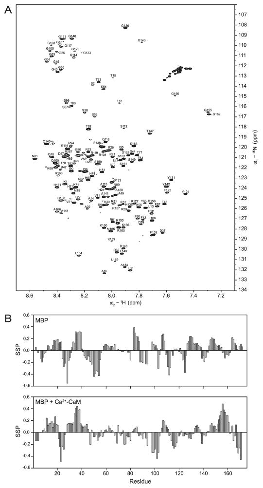
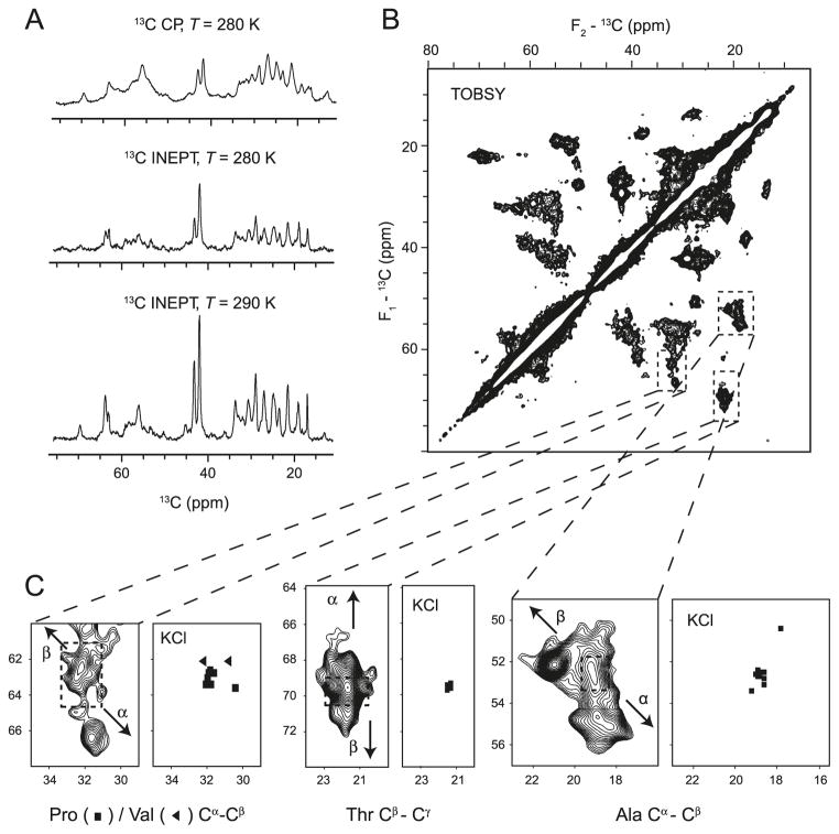
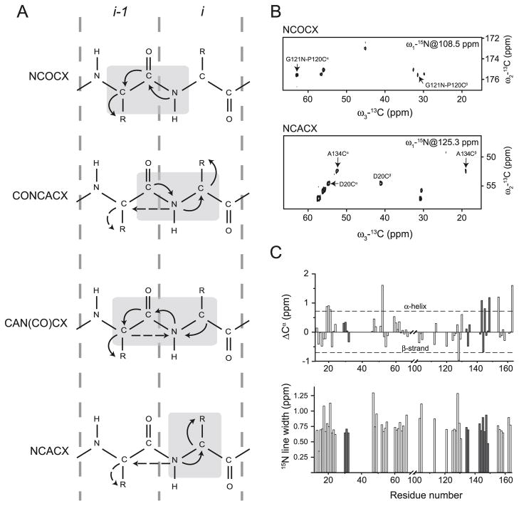
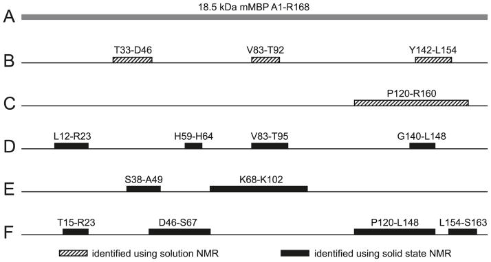
The classic 18.5 kDa isoform of myelin basic protein (MBP) is central to maintaining the structural homeostasis of the myelin sheath of the central nervous system. It is an intrinsically disordered, promiscuous, multifunctional, peripheral membrane protein, whose conformation adapts to its particular environment. Its study requires the selective and complementary application of diverse approaches, of which solution and solid-state NMR spectroscopy are the most powerful to elucidate site-specific features. We review here several recent solution and solid-state NMR spectroscopic studies of 18.5 kDa MBP, and the induced partial disorder-to-order transitions that it has been demonstrated to undergo when complexed with calmodulin, actin, and phospholipid membranes.
Figures
Similar articles
-
Induced secondary structure and polymorphism in an intrinsically disordered structural linker of the CNS: solid-state NMR and FTIR spectroscopy of myelin basic protein bound to actin.Biophys J. 2009 Jan;96(1):180-91. doi: 10.1016/j.bpj.2008.10.003. Biophys J. 2009. PMID: 19134474 Free PMC article.
-
The classic basic protein of myelin--conserved structural motifs and the dynamic molecular barcode involved in membrane adhesion and protein-protein interactions.Curr Protein Pept Sci. 2009 Jun;10(3):196-215. doi: 10.2174/138920309788452218. Curr Protein Pept Sci. 2009. PMID: 19519451 Review.
-
Classic 18.5- and 21.5-kDa myelin basic protein isoforms associate with cytoskeletal and SH3-domain proteins in the immortalized N19-oligodendroglial cell line stimulated by phorbol ester and IGF-1.Neurochem Res. 2012 Jun;37(6):1277-95. doi: 10.1007/s11064-011-0700-2. Epub 2012 Jan 17. Neurochem Res. 2012. PMID: 22249765 Free PMC article.
-
Solid-state NMR spectroscopy of 18.5 kDa myelin basic protein reconstituted with lipid vesicles: spectroscopic characterisation and spectral assignments of solvent-exposed protein fragments.Biochim Biophys Acta. 2007 Dec;1768(12):3193-205. doi: 10.1016/j.bbamem.2007.08.013. Epub 2007 Aug 24. Biochim Biophys Acta. 2007. PMID: 17920035 Free PMC article.
-
MyelStones: the executive roles of myelin basic protein in myelin assembly and destabilization in multiple sclerosis.Biochem J. 2015 Nov 15;472(1):17-32. doi: 10.1042/BJ20150710. Biochem J. 2015. PMID: 26518750 Review.
Cited by
-
The magic of bicelles lights up membrane protein structure.Chem Rev. 2012 Nov 14;112(11):6054-74. doi: 10.1021/cr300061w. Epub 2012 Aug 24. Chem Rev. 2012. PMID: 22920148 Free PMC article. Review. No abstract available.
-
Myelin management by the 18.5-kDa and 21.5-kDa classic myelin basic protein isoforms.J Neurochem. 2013 May;125(3):334-61. doi: 10.1111/jnc.12195. Epub 2013 Mar 6. J Neurochem. 2013. PMID: 23398367 Free PMC article. Review.
-
Nucleus-localized 21.5-kDa myelin basic protein promotes oligodendrocyte proliferation and enhances neurite outgrowth in coculture, unlike the plasma membrane-associated 18.5-kDa isoform.J Neurosci Res. 2013 Mar;91(3):349-62. doi: 10.1002/jnr.23166. Epub 2012 Nov 27. J Neurosci Res. 2013. PMID: 23184356 Free PMC article.
-
Membrane Association Landscape of Myelin Basic Protein Portrays Formation of the Myelin Major Dense Line.Sci Rep. 2017 Jul 10;7(1):4974. doi: 10.1038/s41598-017-05364-3. Sci Rep. 2017. PMID: 28694532 Free PMC article.
-
The proline-rich region of 18.5 kDa myelin basic protein binds to the SH3-domain of Fyn tyrosine kinase with the aid of an upstream segment to form a dynamic complex in vitro.Biosci Rep. 2014 Dec 8;34(6):e00157. doi: 10.1042/BSR20140149. Biosci Rep. 2014. PMID: 25343306 Free PMC article.
References
-
- Ahmed MA, Bamm VV, Shi L, Steiner-Mosonyi M, Dawson JF, Brown L, et al. Induced secondary structure and polymorphism in an intrinsically disordered structural linker of the cns: solid-state NMR and FTIR spectroscopy of myelin basic protein bound to actin. Biophys J. 2009;96(1):180–191. doi: 10.1016/j.bpj.2008.10.003. - DOI - PMC - PubMed
-
- Andrew ER, Bradbury A, Eades RG. Nuclear magnetic resonance spectra from a crystal rotated at high speed. Nature. 1958;182(4650):1659–1659. doi: 10.1038/1821659a0. - DOI
Publication types
MeSH terms
Substances
Grants and funding
LinkOut - more resources
Full Text Sources
Miscellaneous
