. 2010 Apr 29;5(4):e10382.
doi: 10.1371/journal.pone.0010382.

WNT4/beta-catenin pathway maintains female germ cell survival by inhibiting activin betaB in the mouse fetal ovary

Affiliations

WNT4/beta-catenin pathway maintains female germ cell survival by inhibiting activin betaB in the mouse fetal ovary

Chia-Feng Liu et al. PLoS One. .

Abstract

Female germ cells are essential for organogenesis of the ovary; without them, ovarian follicles do not form and functional and structural characteristics of the ovary are lost. We and others showed previously that when either Wnt4 or beta-catenin was inactivated in the fetal ovary, female germ cells underwent degeneration. In this study, we set out to understand whether these two factors belong to the same pathway and how they maintain female germ cell survival. We found that activation of beta-catenin in somatic cells in the Wnt4 knockout ovary restored germ cell numbers, placing beta-catenin downstream of WNT4. In the absence of Wnt4 or beta-catenin, female germ cells entered meiosis properly; however, they underwent apoptosis afterwards. Activin betaB (Inhbb), a subunit of activins, was upregulated in the Wnt4 and beta-catenin knockout ovaries, suggesting that Inhbb could be the cause for the loss of female germ cells, which are positive for activin receptors. Indeed, removal of Inhbb in the Wnt4 knockout ovaries prevented female germ cells from undergoing degeneration. We conclude that WNT4 maintains female germ cell survival by inhibiting Inhbb expression via beta-catenin in the somatic cells. Maintenance of female germ cells hinge upon a delicate balance between positive (WNT4 and beta-catenin) and negative (activin betaB) regulators derived from the somatic cells in the fetal ovary.


Conflict of interest statement

Competing Interests: The authors have declared that no competing interests exist.

Figures

Figure 1
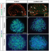
Figure 1. Effects of somatic cell ablation of β-catenin on germ cell apoptosis and entry into meiosis.
(A–B) Immunohistochemistry for germ cell marker TRA98 (red) and apoptotic marker cleaved caspase 3 (green) in control (SF1/cre;Ctnnb1f/+) and β-catenin conditional KO (cKO; SF1/cre;Ctnnb1f/−) ovaries at 17.5 dpc. The arrows indicate cells that are double-positive (yellow) for TRA98 and cleaved caspase 3. The inset in B is an image of a higher magnification of cells double-positive (yellow) for TRA98 and cleaved caspase 3. (C–F) Analysis of the meiotic status of germ cells via immunohistochemistry for SCP3 on germ cell smear obtained from the control and β-catenin conditional KO ovaries at 15.5 dpc. The samples were counterstained with nuclear DAPI (blue). Scale bar represent 100 µm in A–B and 10 µm in C–F.
Figure 2
Figure 2. Effects of constitutively active form of β-catenin on female germ cell survival in the Wnt4 KO ovary.
(A–D) Whole mount light field images of Wnt4 +/−;SF1/cre, Wnt4 −/−;SF1/cre, Wnt4+/−;SF1/cre; Ctnnb1fl.(ex3), and Wnt4−/−;SF1/cre; Ctnnb1fl.(ex3) ovaries at birth. (E) Average total germ cell number was obtained from Wnt4+/−;SF1/cre, Wnt4−/−;SF1/cre, Wnt4+/−;SF1/cre; Ctnnb1fl.(ex3), and Wnt4−/−;SF1/cre; Ctnnb1fl.(ex3 ) ovaries at birth (n = 3 embryos for each genotype). Tukey tests revealed that the average germ cell number in Wnt4−/−;SF1/cre was significantly different from that in Wnt4 +/−;SF1/cre (P = 0.017), Wnt4+/−;SF1/cre; Ctnnb1fl.(ex3) (P = 0.002), and Wnt4−/−;SF1/cre; Ctnnb1fl.(ex3) (P = 0.023). The asterisk represents statistical significance. (F–I) Immunohistochemical staining for androgen-producing enzyme CYP17 (green) was performed in Wnt4 +/−;SF1/cre, Wnt4 −/−;SF1/cre, Wnt4+/−;SF1/cre; Ctnnb1fl.(ex3), and Wnt4−/−;SF1/cre; Ctnnb1fl.(ex3) ovaries at birth. The inset in G represents a higher magnification of the CYP17-positive cells. Scale bar = 100 µm.
Figure 3
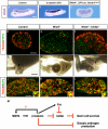
Figure 3. Effects of flutamide treatment on development of the reproductive tract and germ cells in β-catenin conditional KO female.
(A–H) Whole mount images of reproductive tracts and ovaries and (I–L) immunohistochemistry for TRA98 were performed on the ovary of the control (SF1/cre;Ctnnb1f/+) and β-catenin cKO (SF1/cre;Ctnnb1f/−) female with or without flutamide treatment. o =  ovary; arrow =  epididymis, arrowhead =  oviduct. Scale bar represents 500 µm in A–D and 100 µm in E–L.
Figure 4
Figure 4. Involvement of Inhbb in germ cell loss in the absence of Wnt4.
(A–C) Whole mount in situ hybridization for Inhbb was performed on control ovary (SF1/cre;Ctnnbf/+ or Ctnnb1f/−) (A), β-catenin cKO ovary (SF1/cre;Ctnnbf/−) (B), and Wnt4−/−;SF1/cre; Ctnnb1fl.(ex3) ovary (C) at 13.5 dpc. (n = 2–3 for each genotypes). o =  ovary, m =  mesonephros. (D–F) Immunohistochemistry for TRA98 (red) and CYP17 (green) was performed on ovary sections from control (Wnt4+/−; Inhbb+/−), Wnt4 single KO and Wnt4; Inhbb double KO ovary at birth. (G) Light field microscopic images of the reproductive tract were taken from control female, Wnt4 single KO, and Wnt4; Inhbb double KO females at birth. Arrowheads indicate oviduct and arrows indicate epididymal structure. o = ovary. (J–L) Immunohistochemistry for TRA98 (red) and SCP3 (green) were performed on ovary sections from control, Wnt4 single KO and Wnt4; Inhbb double KO ovary at 15.5 dpc. At this stage, the female germ cells have not lost yet in the Wnt4 KO ovaries, allowing us to monitor the status of meiosis. The insets are the images of a higher magnification of cells double-positive (yellow) for TRA98 and SCP3. Scale bar represent 250 µm in A–C, G–I and 100 µm in D–F, J–L. (M) A proposed model for the somatic cell-derived pathway on female germ cell survival: In the mouse fetal ovary, WNT4 signals via β-catenin to decrease the expression of activin βB or Inhbb, which causes loss of meiotic germ cell. WNT4 also stimulates the production of follistatin (Fst), which acts to antagonize the activity of Inhbb. The WNT4/β-catenin pathway also prevents the ectopic production of androgens in the fetal ovary, which is not responsible for the germ cell loss. WNT4 could possibly regulate its own expression via β-catenin.

Similar articles

Cited by

References

    1. Ruggiu M, Speed R, Taggart M, McKay SJ, Kilanowski F, et al. The mouse Dazla gene encodes a cytoplasmic protein essential for gametogenesis. Nature. 1997;389:73–77. - PubMed
    1. McNeilly JR, Saunders PT, Taggart M, Cranfield M, Cooke HJ, et al. Loss of oocytes in Dazl knockout mice results in maintained ovarian steroidogenic function but altered gonadotropin secretion in adult animals. Endocrinology. 2000;141:4284–4294. - PubMed
    1. Soyal SM, Amleh A, Dean J. FIGalpha, a germ cell-specific transcription factor required for ovarian follicle formation. Development. 2000;127:4645–4654. - PubMed
    1. Hashimoto N, Kubokawa R, Yamazaki K, Noguchi M, Kato Y. Germ cell deficiency causes testis cord differentiation in reconstituted mouse fetal ovaries. J Exp Zool. 1990;253:61–70. - PubMed
    1. Behringer RR, Cate RL, Froelick GJ, Palmiter RD, Brinster RL. Abnormal sexual development in transgenic mice chronically expressing mullerian inhibiting substance. Nature. 1990;345:167–170. - PubMed

Publication types