

Silibinin suppresses growth of human colorectal carcinoma SW480 cells in culture and xenograft through down-regulation of beta-catenin-dependent signaling
- PMID: 20454513
- PMCID: PMC2864479
- DOI: 10.1593/neo.10188
Silibinin suppresses growth of human colorectal carcinoma SW480 cells in culture and xenograft through down-regulation of beta-catenin-dependent signaling
Abstract
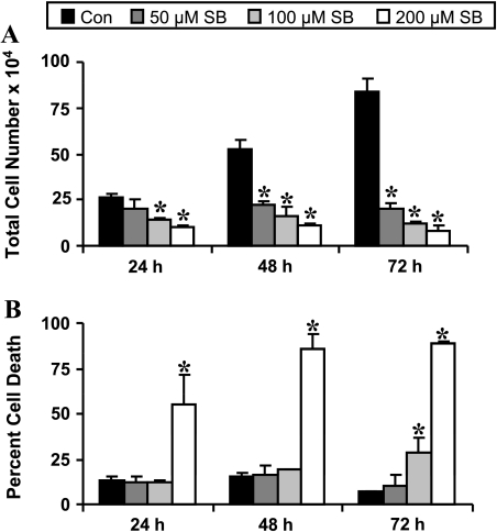
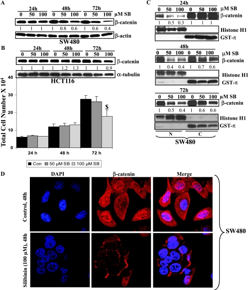
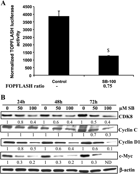
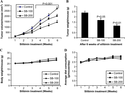
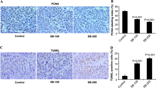
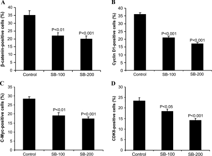
Mutations in APC/beta-catenin resulting in an aberrant activation of Wnt/beta-catenin pathway are common in colorectal cancer (CRC), suggesting that targeting the beta-catenin pathway with chemopreventive/anticancer agents could be a potential translational approach to control CRC. Using human CRC cell lines harboring mutant (SW480) versus wildtype (HCT116) APC gene and alteration in beta-catenin pathway, herein we performed both in vitro and in vivo studies to examine for the first time whether silibinin targets beta-catenin pathway in its efficacy against CRC. Silibinin treatment inhibited cell growth, induced cell death, and decreased nuclear and cytoplasmic levels of beta-catenin in SW480 but not in HCT116 cells, suggesting its selective effect on the beta-catenin pathway and associated biologic responses. Other studies, therefore, were performed only in SW480 cells where silibinin significantly decreased beta-catenin-dependent T-cell factor-4 (TCF-4) transcriptional activity and protein expression of beta-catenin target genes such as c-Myc and cyclin D1. Silibinin also decreased cyclin-dependent kinase 8 (CDK8), a CRC oncoprotein that positively regulates beta-catenin activity, and cyclin C expression. In a SW480 tumor xenograft study, 100- and 200-mg/kg doses of silibinin feeding for 6 weeks inhibited tumor growth by 26% to 46% (P < .001). Analyses of xenografts showed that similar to cell culture findings, silibinin decreases proliferation and expression of beta-catenin, cyclin D1, c-Myc, and CDK8 but induces apoptosis in vivo. Together, these findings suggest that silibinin inhibits the growth of SW480 tumors carrying the mutant APC gene by down-regulating CDK8 and beta-catenin signaling and, therefore, could be an effective agent against CRC.
Figures
Similar articles
-
Histone Demethylase JMJD2D Interacts With β-Catenin to Induce Transcription and Activate Colorectal Cancer Cell Proliferation and Tumor Growth in Mice.Gastroenterology. 2019 Mar;156(4):1112-1126. doi: 10.1053/j.gastro.2018.11.036. Epub 2018 Nov 23. Gastroenterology. 2019. PMID: 30472235
-
Effect of silibinin in human colorectal cancer cells: targeting the activation of NF-κB signaling.Mol Carcinog. 2013 Mar;52(3):195-206. doi: 10.1002/mc.21843. Epub 2011 Nov 15. Mol Carcinog. 2013. PMID: 22086675 Free PMC article.
-
Silibinin inhibits colorectal cancer growth by inhibiting tumor cell proliferation and angiogenesis.Cancer Res. 2008 Mar 15;68(6):2043-50. doi: 10.1158/0008-5472.CAN-07-6247. Cancer Res. 2008. PMID: 18339887
-
Wogonin induced G1 cell cycle arrest by regulating Wnt/β-catenin signaling pathway and inactivating CDK8 in human colorectal cancer carcinoma cells.Toxicology. 2013 Oct 4;312:36-47. doi: 10.1016/j.tox.2013.07.013. Epub 2013 Jul 30. Toxicology. 2013. PMID: 23907061 Review.
-
Revving the Throttle on an oncogene: CDK8 takes the driver seat.Cancer Res. 2009 Oct 15;69(20):7899-901. doi: 10.1158/0008-5472.CAN-09-1704. Epub 2009 Oct 6. Cancer Res. 2009. PMID: 19808961 Free PMC article. Review.
Cited by
-
Developing a high-performance liquid chromatography fast and accurate method for quantification of silibinin.BMC Res Notes. 2019 Nov 14;12(1):743. doi: 10.1186/s13104-019-4774-2. BMC Res Notes. 2019. PMID: 31727143 Free PMC article.
-
The Chalcone Lonchocarpin Inhibits Wnt/β-Catenin Signaling and Suppresses Colorectal Cancer Proliferation.Cancers (Basel). 2019 Dec 7;11(12):1968. doi: 10.3390/cancers11121968. Cancers (Basel). 2019. PMID: 31817828 Free PMC article.
-
MicroRNA-101 is a potential prognostic indicator of laryngeal squamous cell carcinoma and modulates CDK8.J Transl Med. 2015 Aug 19;13:271. doi: 10.1186/s12967-015-0626-6. J Transl Med. 2015. PMID: 26286725 Free PMC article.
-
Nature-derived compounds modulating Wnt/ β -catenin pathway: a preventive and therapeutic opportunity in neoplastic diseases.Acta Pharm Sin B. 2020 Oct;10(10):1814-1834. doi: 10.1016/j.apsb.2019.12.019. Epub 2020 Jan 7. Acta Pharm Sin B. 2020. PMID: 33163337 Free PMC article. Review.
-
Inhibition of Wnt Signaling by Silymarin in Human Colorectal Cancer Cells.Biomol Ther (Seoul). 2016 Jul 1;24(4):380-6. doi: 10.4062/biomolther.2015.154. Epub 2016 Apr 11. Biomol Ther (Seoul). 2016. PMID: 27068260 Free PMC article.
References
-
- Jemal A, Siegel R, Ward E, Hao Y, Xu J, Thun MJ. Cancer statistics, 2009. CA Cancer J Clin. 2009;59:225–249. - PubMed
-
- Mandel JS, Church TR, Bond JH, Ederer F, Geisser MS, Mongin SJ, Snover DC, Schuman LM. The effect of fecal occult-blood screening on the incidence of colorectal cancer. N Engl J Med. 2000;343:1603–1607. - PubMed
-
- Espey DK, Wu XC, Swan J, Wiggins C, Jim MA, Ward E, Wingo PA, Howe HL, Ries LA, Miller BA, et al. Annual report to the nation on the status of cancer, 1975–2004, featuring cancer in American Indians and Alaska Natives. Cancer. 2007;110:2119–2152. - PubMed
-
- Wilkins T, Reynolds PL. Colorectal cancer: a summary of the evidence for screening and prevention. Am Fam Physician. 2008;78:1385–1392. - PubMed
-
- Half E, Arber N. Colon cancer: preventive agents and the present status of chemoprevention. Expert Opin Pharmacother. 2009;10:211–219. - PubMed
Publication types
MeSH terms
Substances
Grants and funding
LinkOut - more resources
Full Text Sources
Other Literature Sources
Medical
Research Materials