. 2010 May 19;22(19):194116.
doi: 10.1088/0953-8984/22/19/194116.

How deeply cells feel: methods for thin gels

Affiliations

How deeply cells feel: methods for thin gels

Amnon Buxboim et al. J Phys Condens Matter. .

Abstract

Tissue cells lack the ability to see or hear but have evolved mechanisms to feel into their surroundings and sense a collective stiffness. A cell can even sense the effective stiffness of rigid objects that are not in direct cellular contact - like the proverbial princess who feels a pea placed beneath soft mattresses. How deeply a cell feels into a matrix can be measured by assessing cell responses on a controlled series of thin and elastic gels that are affixed to a rigid substrate. Gel elasticity E is readily varied with polymer concentrations of now-standard polyacrylamide hydrogels, but to eliminate wrinkling and detachment of thin gels from an underlying glass coverslip, vinyl groups are bonded to the glass before polymerization. Gel thickness is nominally specified using micron-scale beads that act as spacers, but gels swell after polymerization as measured by z-section, confocal microscopy of fluorescent gels. Atomic force microscopy (AFM) is used to measure E at gel surfaces, employing stresses and strains that are typically generated by cells and yielding values for E that span a broad range of tissue microenvironments. To illustrate cell sensitivities to a series of thin-to-thick gels, the adhesive spreading of mesenchymal stem cells was measured on gel mimics of a very soft tissue (eg. brain, E ~ 1 kPa). Initial results show that cells increasingly respond to the rigidity of an underlying 'hidden' surface starting at about 10-20 microm gel thickness with a characteristic tactile length of less than about 5 microm.


Figures
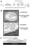
Figure 1
Figure 1
Tissue microenvironments and models. (A) Cellular microenvironments within tissues are characterized by their elasticity E, which ranges over two decades. (B) Anatomically correct, re-traced schematics of mesenchymal microenvironments(–16). Within cartilage (top), chondron units consist of chondrocytes that are embedded within a pericelluler matrix surrounded by a stiff collagenous matrix. Bone-generating osteoblasts (bottom) adhere to a thin and compliant osteoid ECM that is layered on top of rigid calcified bone. (C) Heterogeneous culture models in which a thin and soft matrix is affixed to a rigid substrate.
Figure 2
Figure 2
Surface functionalization of glass substrates for covalent binding of polyacrylamide gels. (A) (1) Cleaned glass substrates (see methods) were silanized with allyltrichlorosilane (ATCS) that forms a dense layer of surface vinyl groups. Covalent attachment of thin polyacrylamide (PA) gels is achieved by direct gel polymerization (2). (B) Irreversible gel attachment to an ATCS treated glass substrate was functionally tested by immersion in ethanol (leading to opaque films) and comparing to a gel that was attached by the standard gluteraldehyde method (GA)(12). Within 30 min, gel detached from GA-treated substrate and is held on a spatula, while the ATCS-immobilized gels remained attached and proved scratch resistant.
Figure 3
Figure 3
Rheological characterization of PA gels. (A) Elasticity of PA gels was measured on a rheometer during gelation. Gel elasticity was controlled by varying acrylamide concentration from 3% to 6% w/v. Black dashed curve connects points of half-maximum stiffness, which illustrates faster polymerization in denser gels. (B) Cylindrical PA gels were stretched and simultaneously imaged to determine strain and relative width (λ*). PA gels in air get thinner with stretch as (strain+1)1/2 as expected for an incompressible material, implying a Poisson’s ratio of 1/2 (black points, dashed line). For comparison, a compressible gel with (strain+1)0.3 is shown as the solid line, and we also show data for networks of the ECM protein fibrin (gray points)(26), with a width that decreases much faster with strain – corresponding to a negative compressibility that reflects protein unfolding.
Figure 4
Figure 4
Preparation of thin PA gels. (A) Thin PA gels were polymerized between an ATCS-treated coverslip and a clean coverslip. To control gel thickness, micron-scale bead spacers were included in the gels and pressed between the glass substrates using a weight. (B) Cross-sections of FITC-labeled gels were obtained using a laser scanning confocal microscope. The red grid indicates 2µm separation. (C) Gel thickness was evaluated using an edge detector, based on the peak centers of the z-dervative (right). (D) Fluorescence intensity profiles of a thick and soft gel were obtained by top-down and bottom-up laser scanning. Intensity profiles show decreasing intensity towards the gel top surface that becomes more prominent for the latter.
Figure 5
Figure 5
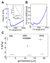
Apparent elasticity of thin gels as evaluated from force-indentation analyses with AFM. (A) Force-indentation curves were fitted (inset) by a variant of the classical Hertz model adjusted for pyramidal (cone-like) tip geometry. (B) E was estimated in the constant-force regime which was robust to changes in fitting range (shown here between 500 nm and 1 µm). (C) The µ-elasticity of thin gels was higher than for intermediate thickness for both soft and stiff gels, indicative of the effective stiffening by the bottom rigid surface and with increased swelling of the intermediate-thickness gels.
Figure 6
Figure 6
How deeply do cells feel? (A) Mesenchymal stem cells were cultured on soft brain-like PA gels of varying thickness. The limit of zero gel thickness while maintain surface chemistry was mimicked using stiff (~34 kPa) collagen-coated PA gels, which have been shown to drive cell morphologies similar to collagen-coated rigid glass (red symbol). Cells were fixed after 24 hr in culture and labeled for actin (red) and DNA (blue). Cell spread area increased with decreasing gel thickness (B) Gel thickness at which MSCs begin to respond to the rigidity of the underlying glass substrate was estimated by a hyperbolic fit of the cell spread area. Compared to an exponential fit, the data was fit better with a hyperbolic relation, which yields 3.4 µm for how far cells feel(31).

Similar articles

Cited by

References

    1. Flanagan LA, Ju YE, Marg B, Osterfield M, Janmey PA. Neurite branching on deformable substrates. Neuroreport. 2002 Dec 20;13(18):2411–2415. - PMC - PubMed
    1. Georges PC, Miller WJ, Meaney DF, Sawyer ES, Janmey PA. Matrices with compliance comparable to that of brain tissue select neuronal over glial growth in mixed cortical cultures. Biophysical Journal. 2006 Apr;90(8):3012–3018. - PMC - PubMed
    1. Patel PN, Smith CK, Patrick CW. Rheological and recovery properties of poly(ethylene glycol) diacrylate hydrogels and human adipose tissue. Journal of Biomedical Materials Research Part A. 2005 Jun 1;73A(3):313–319. - PubMed
    1. Ferrari G, Cusella-De Angelis G, Coletta M, Paolucci E, Stornaiuolo A, Cossu G, et al. Muscle regeneration by bone marrow derived myogenic progenitors. Science. 1998 Mar 6;279(5356):1528–1530. - PubMed
    1. Engler AJ, Griffin MA, Sen S, Bonnetnann CG, Sweeney HL, Discher DE. Myotubes differentiate optimally on substrates with tissue-like stiffness: pathological implications for soft or stiff microenvironments. Journal of Cell Biology. 2004 Sep 13;166(6):877–887. - PMC - PubMed

Publication types