. 2010 May 4;5(5):e10474.
doi: 10.1371/journal.pone.0010474.

Secreted Mycobacterium tuberculosis Rv3654c and Rv3655c proteins participate in the suppression of macrophage apoptosis

Affiliations

Secreted Mycobacterium tuberculosis Rv3654c and Rv3655c proteins participate in the suppression of macrophage apoptosis

Lia Danelishvili et al. PLoS One. .

Abstract

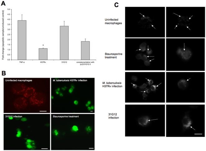
Background: Inhibition of macrophage apoptosis by Mycobacterium tuberculosis has been proposed as one of the virulence mechanisms whereby the pathogen avoids the host defense. The mechanisms by which M. tuberculosis H37Rv strain suppress apoptosis and escapes human macrophage killing was investigated.

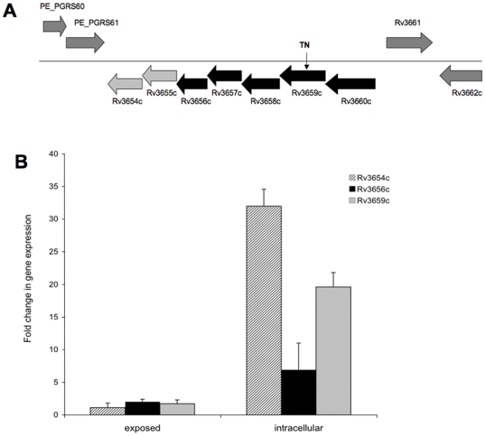
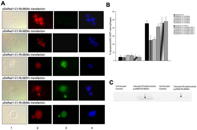
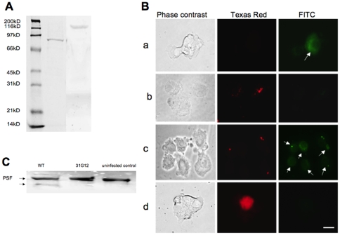
Methodology/principal findings: The screening of a transposon mutant bank identified several mutants, which, in contrast to the wild-type bacterium, had impaired ability to inhibit apoptosis of macrophages. Among the identified genes, Rv3659c (31G12 mutant) belongs to an operon reminiscent of type IV pili. The Rv3654c and Rv3655c putative proteins in a seven-gene operon are secreted into the macrophage cytoplasm and suppress apoptosis by blocking the extrinsic pathway. The operon is highly expressed when the bacterium is within macrophages, compared to the expression level in the extracellular environment. Rv3654c recognizes the polypyrimidine tract binding Protein-associated Splicing Factor (PSF) and cleaves it, diminishing the availability of caspase-8. While M. tuberculosis inhibits apoptosis by the extrinsic pathway, the pathogen does not appear to affect the intrinsic pathway. Inactivation of the intrinsic pathway by pharmacologic agents afftects M. tuberculosis and induces cell necrosis. Likewise, inactivation of PSF by siRNA significantly decreased the level of caspase-8 in macrophages.

Conclusion: While M. tuberculosis inhibits the extrinsic pathway of apoptosis, it appears to activate the intrinsic pathway leading to macrophage necrosis as a potential exit strategy.


Conflict of interest statement

Competing Interests: The authors have declared that no competing interests exist.

Figures

Figure 1
Figure 1. Transposon-inactivated region in the bacterial chromosome.
A Gene organization of 31G12 mutant associated with induction of macrophage apoptosis. The operon contains seven genes, including four genes (Rv3657c–Rv3660c) that resemble a pilus assembly proteins, type II/IV secretion system proteins, as well as tight adherence genes of TadZ, TadA-C, respectively; Rv3656c transmembrane protein and Rv3654c, Rv3655c genes that encode secreted proteins. The arrow indicates the gene disrupted by the insertion of Tn5367 transposon (TN). B M. tuberculosis gene expression following 30 min exposure to macrophages or 3 h infection. Total RNAs from broth-grown bacteria, as well as from intracellular- and macrophage-exposed bacteria, were used to determine the copy numbers of cDNAs for target and reference genes. Data were analyzed on Ct values basis for each sample and normalized with an internal housekeeping gene control, 16S rRNA.
Figure 2
Figure 2. Effect on apoptotic pathways.
A M. tuberculosis infection prevents caspase-8 activation in macrophages. U937 cells were infected or uninfected and/or treated with recombinant TNF-α protein, and fold change of caspase-8 activation was calculated according the manufacturer's protocol. The *, statistically significant difference between TNF-α-treated and M. tuberculosis-infection groups (p<0.05). B Immunofluorescence analysis of mitochondrial transmembrane disruption after M. tuberculosis H37Rv, 31G12 infection and staurosporine treatment, compared with uninfected macrophages. Red dots indicate undamaged mitochondria in uninfected cells. Bar, 10 µm. C M. tuberculosis infection promotes cytochrome C release from the mitochondria into the cytoplasm of U937 cells. Macrophages were infected with wild-type bacterium or 31G12 mutant and were then stained for cytochrome C. Staurosporine treatment was used as positive control. The arrows on the punctuated staining indicate cytochrome C localization in the mitochondria in uninfected macrophages; diffuse staining shows characteristic mitochondrial cytochrome C release in M. tuberculosis H37Rv- or 31G12-infected cells, similar to the staurosporine treatment. Two images were included for each experimental group. Bar, 10 µm.
Figure 3
Figure 3. Macrophage transfection affects apoptosis.
A CaspaTag™ In Situ analysis of U937 macrophages for caspase-8 activation and nuclear changes during transfection with M. tuberculosis proteins. While transfection with M. tuberculosis Rv3654c and Rv3655c genes during TNF-α treatment of macrophages suppressed caspase-8 activation and nuclear fragmentation, M. tuberculosis Rv3656c-, Rv3657c- and Rv3659c- transfected and TNF-α-treated macrophages showed caspase-8 activation and nuclear morphological changes typical of apoptosis. Colors in the columns: (1) Phase contrast, (2) Red – transfection, (3) Green – caspase-8 activation and (4) Blue – nuclear staining with Hoechst stain. Bar, 10 µm. B Effect of M. tuberculosis-secreted proteins on caspase-8 activation during macrophage apoptosis. U937 cells were transfected with Rv3654c–Rv3659c genes for 8 h and then treated with human recombinant TNF-α protein (0.5 µM). Active caspase-8 was quantified using In Situ Assay kit (Chemicon). C Analysis of secreted proteins in the cytoplasm of infected macrophages. U937 cells were infected with M. tuberculosis containing pJAM2:Rv3654c or pJAM2:Rv3655c over-expressed vectors at an MOI 1∶10. Concentrated bacterial and cell lysate-free supernatants were subjected to Western blotting using His-tag antibody.
Figure 4
Figure 4. His-tagged pull-down analysis.
A His-tagged pull-down assay of cellular proteins interacting with M. tuberculosis Rv3654c and Rv3655c proteins identified 76kDa PSF (1) and 175kDa ALO17 (2), respectively. B PSF protein levels in U937 cells. Macrophages were uninfected (a) or infected with M. tuberculosis wild-type (b); 31G12 mutant (c); and transfected with pLDRv3654c (d). Bacteria (red) were labeled with rhodamine and the PSF protein was visualized by fluorescein conjugated secondary antibody (green). Bar, 10 µm. C Western blot analysis of PSF protein after 24 h of infection with M. tuberculosis wild-type and 31G12 mutant.
Figure 5
Figure 5. Macrophage transfection with siRNA.
(A) PSF protein expression levels by Western blot analysis: 1. Untreated cells, 2. Scrambled siRNA control transfection, 3. PSF siRNA transfection. B PSF protein band intensity levels measured with Li-Cor Odyssey Imaging software. C Caspase-8 protein expression levels by Western blot analysis: 1. Recombinant TNF-α treatment (positive control), 2. Untreated cells, 3. Scrambled siRNA control transfection and recombinant TNF-α treatment, 4. PSF siRNA transfection and recombinant TNF-α treatment. D Caspase-8 protein band intensity levels measured with Li-Cor Odyssey Imaging software.
Figure 6
Figure 6. Macrophage apoptosis and necrosis.
A Comparison of the levels of macrophage apoptosis and necrosis during M. tuberculosis H37Rv infection at later time points. M. tuberculosis induced significantly more necrosis of macrophages (64±0.5%) at day 9 (p<0.05) than apoptosis (18±1.8%), compared with uninfected controls. The decreased level of apoptosis during the infection with virulent M. tuberculosis is related to duration of macrophage infection; however, M. tuberculosis triggers a greater level of necrosis at later time points than apoptosis. B Inhibitory effect of Z-LEHD-FMK.TFA (caspase-9 inhibitor) on necrosis levels of macrophages. Comparisons of M. tuberculosis H37Rv-induced apoptosis and necrosis at different time points did not show significant induction of necrosis after treatment of infected macrophages with caspase-9 inhibitor.

Similar articles

Cited by

References

    1. Ting LM, Kim AC, Cattamanchi A, Ernst JD. Mycobacterium tuberculosis inhibits IFN-gamma transcriptional responses without inhibiting activation of STAT1. J Immunol. 1999;163:3898–3906. - PubMed
    1. Sturgill-Koszycki S, Schlesinger PH, Chakraborty P, Haddix PL, Collins HL, et al. Lack of acidification in Mycobacterium phagosomes produced by exclusion of the vesicular proton-ATPase. Science. 1994;263:678–681. - PubMed
    1. Via LE, Deretic D, Ulmer RJ, Hibler NS, Huber LA, et al. Arrest of mycobacterial phagosome maturation is caused by a block in vesicle fusion between stages controlled by rab5 and rab7. J Biol Chem. 1997;272:13326–13331. - PubMed
    1. Flynn JL, Chan J. Immunology of tuberculosis. Annu Rev Immunol. 2001;19:93–129. - PubMed
    1. Davis AS, Vergne I, Master SS, Kyei GB, Chua J, et al. Mechanism of Inducible Nitric Oxide Synthase Exclusion from Mycobacterial Phagosomes. PLoS Pathog. 2007;3:e186. - PMC - PubMed

Publication types

MeSH terms