. 2010 May 4;5(5):e10458.
doi: 10.1371/journal.pone.0010458.

A novel cancer vaccine strategy based on HLA-A*0201 matched allogeneic plasmacytoid dendritic cells

Affiliations

A novel cancer vaccine strategy based on HLA-A*0201 matched allogeneic plasmacytoid dendritic cells

Caroline Aspord et al. PLoS One. .

Abstract

Background: The development of effective cancer vaccines still remains a challenge. Despite the crucial role of plasmacytoid dendritic cells (pDCs) in anti-tumor responses, their therapeutic potential has not yet been worked out. We explored the relevance of HLA-A*0201 matched allogeneic pDCs as vectors for immunotherapy.

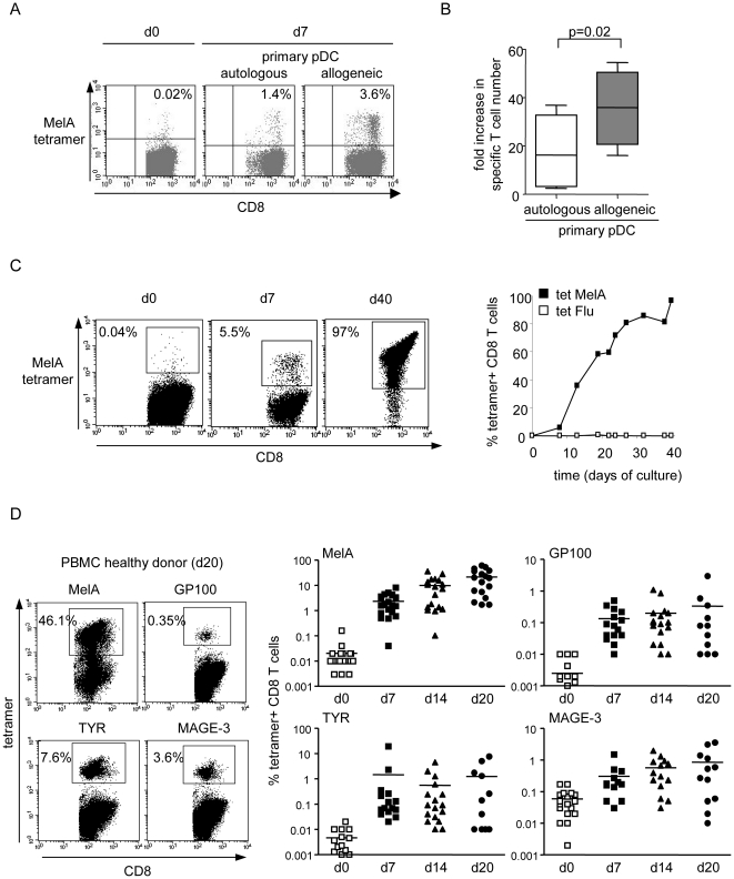
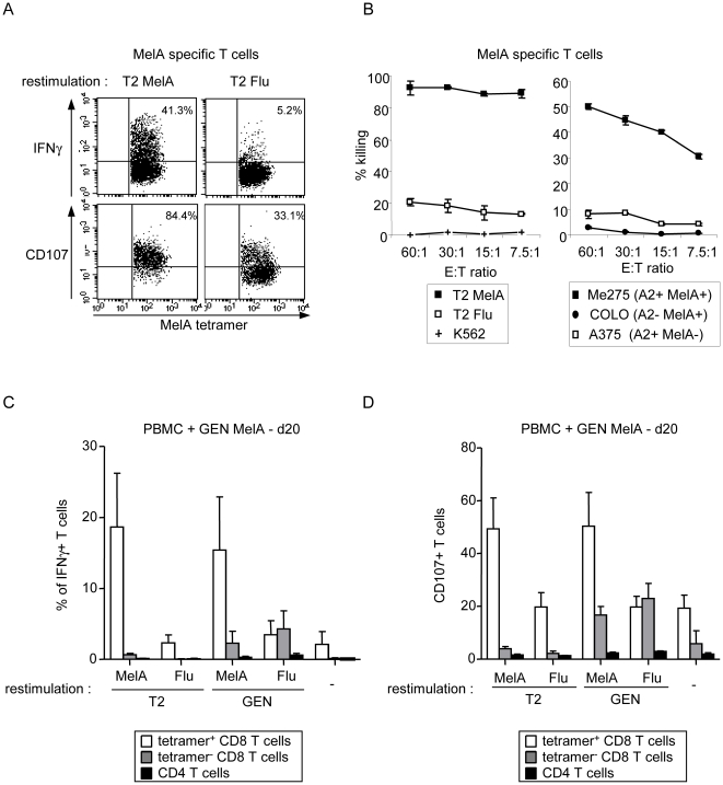
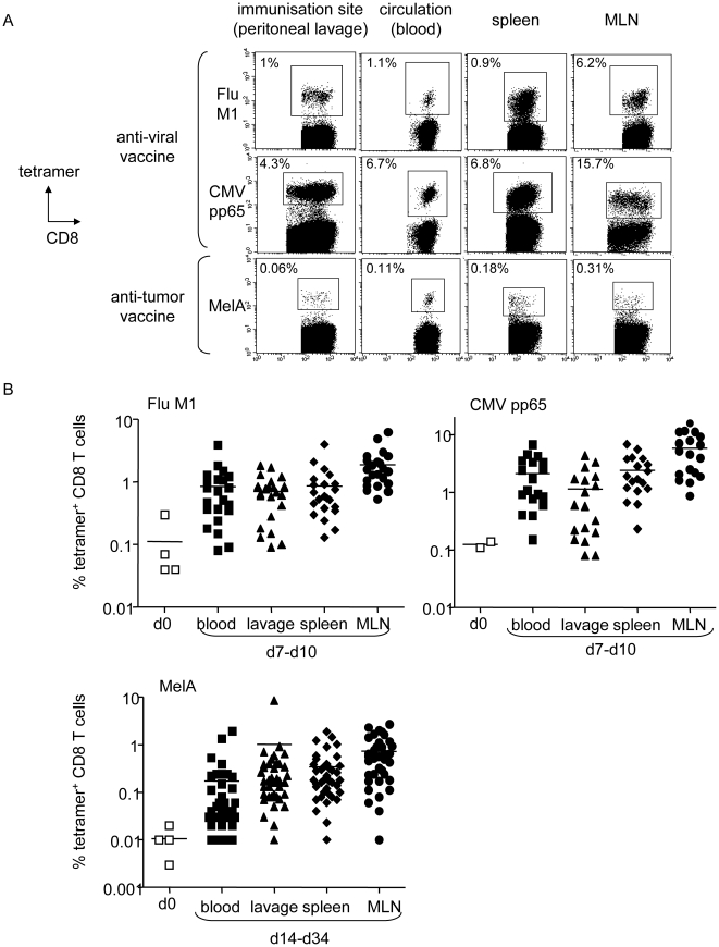
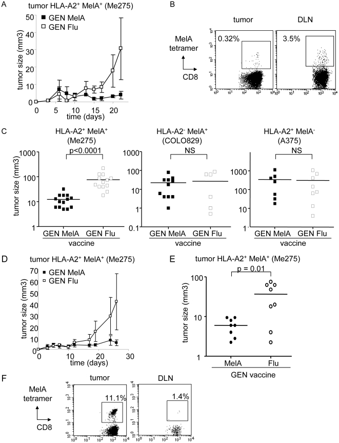
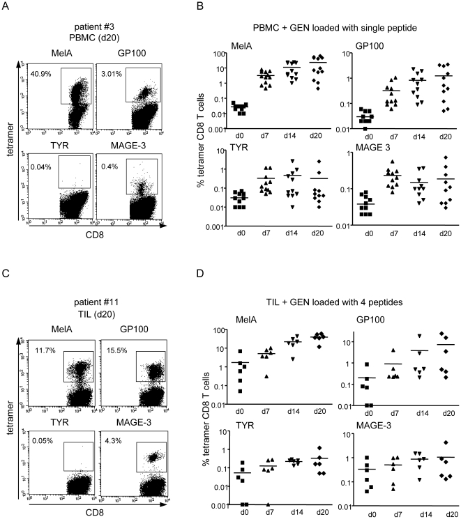
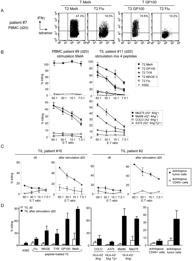
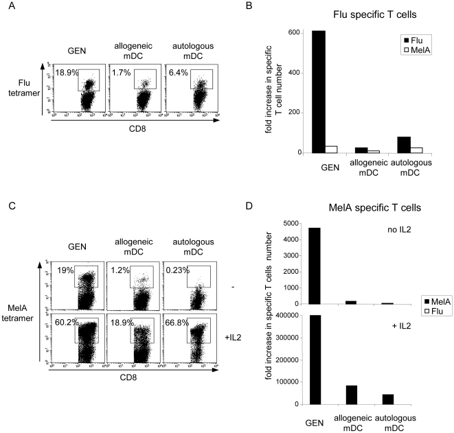
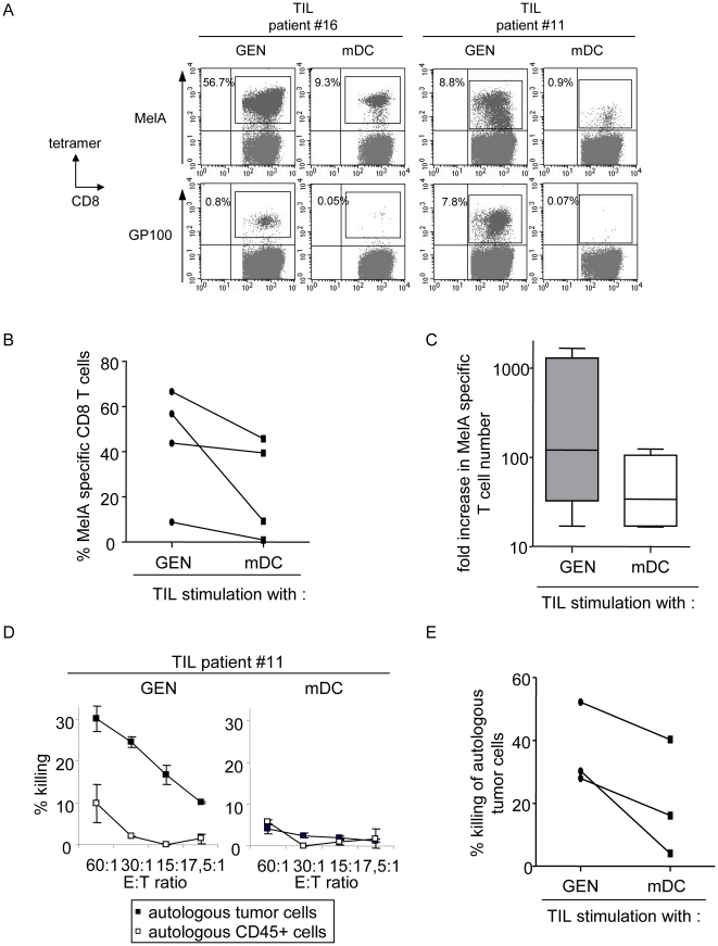
Methods and findings: Stimulation of PBMC from HLA-A*0201(+) donors by HLA-A*0201 matched allogeneic pDCs pulsed with tumor-derived peptides triggered high levels of antigen-specific and functional cytotoxic T cell responses (up to 98% tetramer(+) CD8 T cells). The pDC vaccine demonstrated strong anti-tumor therapeutic in vivo efficacy as shown by the inhibition of tumor growth in a humanized mouse model. It also elicited highly functional tumor-specific T cells ex-vivo from PBMC and TIL of stage I-IV melanoma patients. Responses against MelA, GP100, tyrosinase and MAGE-3 antigens reached tetramer levels up to 62%, 24%, 85% and 4.3% respectively. pDC vaccine-primed T cells specifically killed patients' own autologous melanoma tumor cells. This semi-allogeneic pDC vaccine was more effective than conventional myeloid DC-based vaccines. Furthermore, the pDC vaccine design endows it with a strong potential for clinical application in cancer treatment.

Conclusions: These findings highlight HLA-A*0201 matched allogeneic pDCs as potent inducers of tumor immunity and provide a promising immunotherapeutic strategy to fight cancer.


Conflict of interest statement

Competing Interests: The authors have declared that no competing interests exist.

Figures

Figure 1
Figure 1. HLA-A*0201 matched allogeneic pDCs induce highly effective tumor-specific T cell responses from HLA-A*0201+ healthy donors' PBMC in vitro.
(A,B) Autologous or allogeneic HLA-A*0201+ primary pDC sorted from the blood of healthy donors were pulsed with MelA peptide and used to stimulate HLA-A*0201+ PBMC. The specific T cell response was analyzed at d7 by tetramer labelling. (A) Percentage of MelA specific T cells (gated on CD8+ T cells). One representative experiment is shown. (B) Amplification of the absolute number of specific T cells from d0 to d7 (4 independent experiments performed with 3 different donors). (C,D) allogeneic HLA-A*0201+ PBMC from healthy donors were stimulated with the irradiated peptide-loaded HLA-A*0201+ pDC line and weekly restimulated in the presence of IL2. Specificity of the T cells was determined by tetramer labelling and flow cytometry analysis. (C) Representative dotplots gated on CD8+ T cells (left panel) and percentages (right panel) of MelA tetramer+ T cells in the culture initially and at different time points after stimulation with the pDC line loaded with MelA peptide. Flu tetramer was used as control. (D) Representative dot plots gated on CD8+ T cells (left panel) and percentages of tetramer+ CD8+ T cells obtained at days 7, 14 and 20 of the culture towards MelA (n = 18), GP100 (n = 16), TYR (n = 16) and MAGE-3 (n = 16) tumor antigens.
Figure 2
Figure 2. The tumor-specific T cells primed by the HLA-A*0201 matched allogeneic pDC line in vitro exhibited functional antigen- and HLA-A*0201-specific activity.
(A) MelA-specific T cells induced by the pDC line secrete IFNγ and express CD107 on the surface upon specific restimulation. Cells from the culture (day 14) were submitted to tetramer labelling and restimulated with T2 cells pulsed with a relevant or control peptide. IFNγ production was assessed by intracellular staining and CD107 expression by adding anti-CD107a+b antibodies during the restimulation. Dotplots are gated on tetramer+ CD8+ T cells. Representative of 8 experiments performed with 3 donors at day 8–40 of the culture. (B) MelA-specific T cells induced by the pDC line are cytotoxic. T cells were selected from the culture and submitted to a 51Cr release assay using peptide-loaded T2 cells and melanoma tumor cells as targets. Representative of 8 experiments performed with 4 donors at d13–40 of the culture. (C,D) IFNγ secretion and CD107 expression were assessed as described in (A) after three stimulations of PBMC and analyzed on the tetramer+ CD8+ T cells (white bars) and on the non-specific tetramer- CD8+ T cells (grey bars) and CD4+ T cells (black bars) upon restimulation with peptide-pulsed T2 or GEN cells (4 experiments for each condition).
Figure 3
Figure 3. Vaccination with the peptide-loaded HLA-A*0201 matched allogeneic pDC line elicits strong antigen-specific T cell responses in humanized mice.
(A-B) Immunodeficient NOD-SCIDβ2m-/- mice were reconstituted intraperitoneally with 50.106 human HLA-A*0201+ healthy donors' PBMC and vaccinated by the same route with 5.106 irradiated peptide-loaded GEN cells. Specific T cell induction was analyzed at the injection site (lavage), in the circulation (blood) and lymphoid organs (spleen, LN) by tetramer labelling of human T cells in cell suspensions. (A) Vaccination with peptide-loaded GEN cells induced specific T cell responses in vivo. Representative dot plots of tetramer labeled T cells induced after a single vaccination with peptide-loaded GEN cells in different organs at day 8 for anti-viral vaccine (Flu, CMV) and day 10 for anti-tumor vaccine (MelA) (gated on CD8+ T cells). One mice per group is shown. Initial levels of specific T cells within PBMC were 0.04%, 0.14% and 0.003% respectively. (B) Levels of specific T cells before (day 0) and after vaccination with GEN loaded with FluM1 (n = 22 mice, 4 donors, 1 vaccine), CMVpp65 (n = 18 mice, 2 donors, 1 vaccine) and MelA (n = 38 mice, 4 donors, 2–3 vaccines) peptides at the indicated times in different organs. Each dot represents one vaccinated HuPBL mice (bars at mean).
Figure 4
Figure 4. Vaccination with the peptide-loaded HLA-A*0201 matched allogeneic pDC line protect humanized mice from tumor development both prophylactically and therapeutically.
(A-C) Immunodeficient NOD-SCID β2m-/- mice reconstituted intraperitoneally with human HLA-A*0201+ PBMC (HuPBL mice) were weekly vaccinated subcutaneously with irradiated MelA or Flu-loaded GEN cells and challenged 5 days later with melanoma tumor cells in the flank. (A) Follow up of tumor progression. One experiment representative of 5. (B) Tetramer labelling of tumor and draining LN cell suspensions from HuPBL mice vaccinated with MelA-loaded GEN cells showing the presence of MelA-specific T cells (gated on CD8+ T cells). (C) The therapeutic effects of the vaccine are HLA-A*0201-restricted and antigen-specific. Comparative tumor size 27 days after implantation of Me275, COLO829 and A375 melanoma cells into HuPBL mice vaccinated with MelA or Flu-loaded GEN cells (pool of 3 independent experiments for each tumor type performed with 6 to 14 mice per group). (D-F) Immunodeficient NOD-SCID β2m-/- mice reconstituted intraperitoneally with human HLA-A*0201+ PBMC (HuPBL mice) were first challenged with melanoma Me275 tumor cells in the flank and then vaccinated subcutaneously with irradiated MelA or Flu-loaded GEN cells weekly starting 4 days later. (D) Follow up of tumor progression. One representative experiment out of 2. (E) Comparative tumor size 25 days after tumor implantation (pool of 2 independent experiments, 8 mice/group). (F) Tetramer labelling of tumor and draining LN cell suspensions from HuPBL mice vaccinated with MelA-loaded GEN cells showing the presence of MelA-specific T cells (gated on CD8+ T cells).
Figure 5
Figure 5. The HLA-A*0201 matched allogeneic pDC line loaded with melanoma-derived peptides induces multi-specific T cell responses ex-vivo from stage I-IV melanoma patients.
PBMC (n = 12) and TIL (n = 6) obtained from stage I-IV HLA-A*0201+ melanoma patients were cultured with irradiated GEN cells loaded with MelA, GP100, TYR and/or MAGE-3 derived peptides and restimulated every 7 days. Percentages of specific T cells were determined by tetramer labelling after culture of PBMC with single peptide-loaded GEN cells (A,B) and of TIL with GEN loaded with a mix of the 4 peptides (C,D). Representative experiments with PBMC (A) and TIL (C) are shown at day 20 of the culture. Results from PBMC and TIL cohorts are shown in (B) and (D) at days 0, 7, 14 and 20 of culture. For TYR (D), one patient was excluded due to an extremely intense response (see Figure S5).
Figure 6
Figure 6. The melanoma patients' tumor-specific T cells induced by the HLA-A*0201 matched allogeneic pDC line are highly cytotoxic and lyse autologous melanoma tumor cells.
Tumor-specific T cells induced by the pDC line loaded with melanoma-derived peptides from melanoma patients' PBMC and TIL are highly functional in an HLA-A*0201 and antigen-specific manner. (A) Tetramer+ T cells specifically secreted IFNγ upon restimulation with T2 cells pulsed with the relevant peptide. Representative of 6 PBMC and 2 TIL samples. (B-D) T cells exhibited cytotoxicity towards allogeneic and autologous melanoma tumor cells and relevant peptide-pulsed T2 cells. T cells induced from PBMC or TIL were purified from days 15–20 cultures and used in a 51Cr release assay against peptide-loaded T2 cells, allogeneic and autologous melanoma tumor cells, and autologous CD45+ cells. (B) The PBMC sample shown was stimulated with MelA-loaded GEN cells. The TIL sample shown was stimulated with GEN cells loaded with a mixture of 4 peptides and developed a response towards MelA, GP100 and MAGE-3 antigens (see Figure 5C). Representative of 12 PBMC and 6 TIL samples. (C) Percentage of killing of autologous tumor cells compared to autologous CD45+ cells by TIL before and after stimulation. Representative of 6 TIL samples. (D) Comparison of the killing capacity between unstimulated and stimulated TIL on the indicated targets at a 60∶1 ratio. Mean+/-SEM of 6 TIL samples.
Figure 7
Figure 7. The HLA-A*0201 matched allogeneic pDC line is more effective than conventional mDCs.
PBMC from HLA-A*0201+ healthy donors were stimulated either with the pDC line or with allogeneic or autologous HLA-A*0201+ mDCs loaded with Flu (A, B) or MelA (C, D) peptide in presence of IL-2 where indicated. (A) Percentages of FluM1 tetramer+ CD8+ T cells and (B) fold increase in specific T cell number at d7 of culture. MelA tetramer was used as control. (C) Percentages of MelA tetramer+ CD8+ T cells and (D) fold increase in specific T cell number at d20 of culture. Flu tetramer was used as control.
Figure 8
Figure 8. The HLA-A*0201 matched allogeneic pDCs are more potent at inducing tumor-specific T cells from melanoma patients than conventional mDCs.
TIL from melanoma patients were cultured either with irradiated GEN cells or allogeneic LPS-matured HLA-A*0201+ mDCs loaded with a mixture of MelA, GP100, TYR and MAGE-3 derived peptides. Percentages of tumor-specific T cells were determined by tetramer labelling after three stimulations at day 20 of culture. (A) Representative dotplots of two cultures. (B) Comparative percentages of MelA-specific T cells and (C) fold increase in specific T cell numbers upon stimulation of TIL with GEN or mDCs (4 patients). (D,E) T cells were purified from the culture and submitted to a 51Cr release assay. (D) Percentage of killing of autologous tumor cells and autologous CD45+ cells after TIL stimulation with GEN or mDCs. One representative patient is shown. (E) Comparative killing efficacy against autologous tumor cells at a 60∶1 ratio.

Similar articles

Cited by

References

    1. Finn OJ. Cancer immunology. N Engl J Med. 2008;358:2704–2715. Review. - PubMed
    1. Thurner B, Haendle I, Röder C, Dieckmann D, Keikavoussi P, et al. Vaccination with mage-3A1 peptide-pulsed mature, monocyte-derived dendritic cells expands specific cytotoxic T cells and induces regression of some metastases in advanced stage IV melanoma. J Exp Med. 1999;190:1669–1678. - PMC - PubMed
    1. Palucka AK, Dhodapkar MV, Paczesny S, Ueno H, Fay J, et al. Boosting vaccinations with peptide-pulsed CD34+ progenitor-derived dendritic cells can expand long-lived melanoma peptide-specific CD8+ T cells in patients with metastatic melanoma. J Immunother. 2005;28:158–168. - PubMed
    1. Gattinoni L, Powell DJ, Jr, Rosenberg SA, Restifo NP. Adoptive immunotherapy for cancer: building on success. Nat Rev Immunol. 2006;6:383–393. Review. - PMC - PubMed
    1. Dudley ME, Wunderlich JR, Yang JC, Sherry RM, Topalian SL, et al. Adoptive cell transfer therapy following non-myeloablative but lymphodepleting chemotherapy for the treatment of patients with refractory metastatic melanoma. J Clin Oncol. 2005;23:2346–2357. - PMC - PubMed

Publication types