. 2010 May 4;8(5):e1000365.
doi: 10.1371/journal.pbio.1000365.

Sex and the single cell. II. There is a time and place for sex

Affiliations

Sex and the single cell. II. There is a time and place for sex

Carmen C Robinett et al. PLoS Biol. .

Abstract

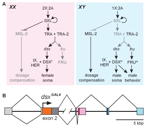
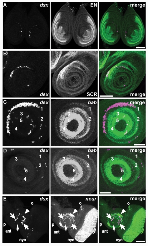
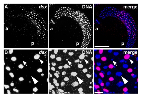
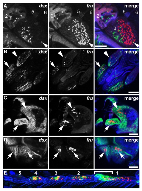
The Drosophila melanogaster sex hierarchy controls sexual differentiation of somatic cells via the activities of the terminal genes in the hierarchy, doublesex (dsx) and fruitless (fru). We have targeted an insertion of GAL4 into the dsx gene, allowing us to visualize dsx-expressing cells in both sexes. Developmentally and as adults, we find that both XX and XY individuals are fine mosaics of cells and tissues that express dsx and/or fruitless (fru(M)), and hence have the potential to sexually differentiate, and those that don't. Evolutionary considerations suggest such a mosaic expression of sexuality is likely to be a property of other animal species having two sexes. These results have also led to a major revision of our view of how sex-specific functions are regulated by the sex hierarchy in flies. Rather than there being a single regulatory event that governs the activities of all downstream sex determination regulatory genes-turning on Sex lethal (Sxl) RNA splicing activity in females while leaving it turned off in males-there are, in addition, elaborate temporal and spatial transcriptional controls on the expression of the terminal regulatory genes, dsx and fru. Thus tissue-specific aspects of sexual development are jointly specified by post-transcriptional control by Sxl and by the transcriptional controls of dsx and fru expression.


Conflict of interest statement

The authors have declared that no competing interests exist.

Figures

Figure 1
Figure 1. The Drosophila sex hierarchy.
(A) The sex determination hierarchy in Drosophila regulates all sexually dimorphic aspects of development. The ratio of X chromosomes to autosomes determines the RNA splicing activity of SXL, which leads to sex-specific alternative splicing of dsx and fru transcripts at the bottom of the hierarchy. The resulting male- and female-specific DSX and FRU isoforms confer sexual identity to the cells in which they are produced. (Adapted from .) (B) The dsxGAL4 gene. Targeted insertion of GAL4 coding sequence (orange box) after the translational start codon (arrow) of dsx in exon 2 allows expression of GAL4 wherever dsx is expressed. Chromosomal sequences are color-coded as follows: 5′ UTR sequences common to the mRNAs of both sexes (light gray), N-terminal coding sequence common to mRNAs of both sexes (dark gray), coding and 3′ UTR sequences specific to females (magenta and pink, respectively), coding and 3′ UTR sequences specific to males (dark and light blue, respectively), and intronic sequences (black line). Male-specific and female-specific splicing patterns are depicted above or below the chromosome, respectively. Slash marks in the second intron represent ∼24 kb of DNA.
Figure 2
Figure 2. dsxGAL4 driving UAS-induced fluorescent reporters recapitulates known patterns of gonadal dsx expression.
(A) dsxGAL4 expression is restricted to cells of the paired uncoalesced gonads in embryonic stage 13. The embryo posterior is marked by an asterisk, and native GFP fluorescence was imaged from the membrane reporter, UAS-mCD8::GFP. Scale bar, 100 µm. (B–C) Expression of dsxGAL4 in the male third instar larval gonad is restricted to somatic cells. Reporter is UAS-RedStinger (nuclear DsRed). (Bi) dsxGAL4-expressing cells include the clustered hub cells of the stem cell niche at the gonad apical tip (arrow), cyst cells dispersed throughout the gonad (barbed arrowhead), and terminal epithelial cells with small round nuclei at the basal end of the gonad (asterisk). A fat body cell nucleus is indicated (arrowhead). (Bii) dsxGAL4 expression (magenta) is excluded from the germline cells marked with cytoplasmic VASA (green). Scale bar, 50 µm. (C) Expression of the JFRC-IVSA membrane-bound GFP reporter (green) reveals sheath cell cytoplasmic processes ramifying through cysts of male germ cells in the basal half of the gonad. DNA of the large male germline cells is stained with DAPI (magenta). Gonad posterior indicated with an asterisk. Single confocal section shown. Scale bar, 20 µm. (D) dsxGAL4 expression in the female third instar larval gonad is restricted to somatic cells (magenta) and is excluded from the germline cells marked with cytoplasmic VASA (green). A fat body cell nucleus is indicated (arrowhead). Reporter is UAS-RedStinger (nuclear DsRed). Scale bar, 50 µm. (E) dsxGAL4 expression in hub cells of the stem cell niche at the apical tip of male the gonad was visualized with the JFRC-IVSA membrane-bound GFP reporter (Eii; green in Eiv). Processes of dsxGAL4-expressing cells enveloped germline cells marked with VASA (Ei; blue in Eiv), including the ring of germline stem cells adjacent to the hub (arrows). Expression in hub cells is confirmed by overlap with DN-Cadherin (DN-Cad) (Eiii; red in Eiv), which marks membranes of the hub cells (arrowheads). Overlap of membrane-bound GFP with DN-Cadherin is yellow in the merge (Eiv). Scale bar, 50 µm.
Figure 3
Figure 3. dsxGAL4-induced expression of UAS-dsxIR at 29°C causes transformation of a sexually dimorphic row of leg bristles toward an intersexual morphology.
(A) Tarsal segment 1 from chromosomal males (XY) and females (XX) with the dsxGAL4 chromosome alone (CyO, balancer chromosome) or driving UAS-dsxIR. The regions of the male sex comb and homologous female last transverse bristle row are bracketed. UAS-dsxIR causes the thickness, pigmentation, and taper of bristles to look the same in males and females. (B) Quantitation of bristle numbers from experiment in (A). Histogram bars for the UAS-dsxIR condition are marked with an asterisk for each chromosomal sex. XY CyO males (n = 17 legs) have an average of 10.2±0.62 (standard deviation) bristles, which is reduced to 8.5±0.61 in XY UAS-dsxIR (n = 19). XX CyO females (n = 19) have an average of 5.35±0.49 bristles, which is increased to 6.47±0.611 in XX UAS-dsxIR (n = 18).
Figure 4
Figure 4. dsxGAL4 is expressed in imaginal discs.
Fluorescence of indicated reporters in imaginal discs of third instar female larvae is shown. (A) dsxGAL4 driving expression of UAS-Stinger nuclear GFP reporter (magenta) is compared to the posterior compartment of the disc marked by EN (green) in this pair of foreleg discs from an early wandering third instar larva. At this stage, few cells express dsxGAL4, but the majority of these cells form a broken crescent within the anterior compartment. Scale bar, 100 µm. (B) Expression of UAS-Stinger nuclear GFP reporter (magenta) in the foreleg disc is compared to the position of tarsal and tibial primordia that express high levels of SCR (green). In this disc from a more mature wandering third instar larva than shown in (A) there are more expressing cells in the dsxGAL4 crescent, and the crescent overlaps with the SCR domain. Scale bar, 100 µm. (C) Expression of UAS-RedStinger nuclear DsRed reporter (magenta) is compared to the position of tarsal segment boundaries defined by concentric expression of bab-lacZ (green). In this foreleg disc from a third instar larva shortly before purarium formation, there is a greater number of cells expressing dsxGAL4 in the dsxGAL4 crescent, and the crescent overlaps with the bab-lacZ domain corresponding to the T1 tarsal segment. Scale bar, 50 µm. (D) Expression of UAS-RedStinger nuclear DsRed reporter (magenta) in a leg disc corresponding to the second or third leg is compared to the position of bab-lacZ (green). In contrast to (A–C), there are only a few cells expressing dsxGAL4 scattered across the disc, and there is no crescent. Scale bar, 50 µm. (E) Expression of UAS-RedStinger nuclear DsRed reporter (magenta) is compared to the position of cells expressing the proneural marker neur-lacZ (green) in the eye-antennal disc from a mature wandering third instar larva. Many cells express dsxGAL4 in the antennal (ant) portion of the disc, while only a few cells express dsxGAL4 in the eye portion. Primordia for the second (arrowhead) and third (barbed arrows) antennal segments are indicated, as are primordia for the arista (arrow), palpus (p), and ocellus (o). Scale bar, 50 µm.
Figure 5
Figure 5. dsxGAL4 is expressed in a subset of cells of the midgut.
(A) The frequency of cells expressing UAS-RedStinger nuclear DsRed reporter (magenta) varies along the length of the anterior midgut from an adult female. Non-expressing nuclei are marked with DAPI alone (blue). Anterior (a) and posterior (p). Speckles of autofluorescent debris are seen in the area outside of the gut. Scale bar, 100 µm. (B) Magnified view of a region in (A). Some enterocytes express dsxGAL4 (small arrowhead) while others do not (large arrowhead). In this region of the gut, cells with smaller nuclei (barbed arrow) that are likely to be enterocrine or intestinal stem cells do not express dsxGAL4. Scale bar, 10 µm.
Figure 6
Figure 6. dsxGAL4 and fruP1.LexA expression in peripheral sensory structures.
Expression of dsxGAL4 and fruP1.LexA at 72 h APF. In the fruP1.LexA chromosome, LexA::VP16 coding sequence followed by the SV40 poly-A signal and α-tubulin 3′UTR sequences as transcriptional termination sequences were inserted into the fru gene . dsxGAL4 was detected by expression of UAS-Stinger nuclear GFP reporter (green), and fruP1.LexA was detected by expression of lexAop-tdTomato::nls (red). Cuticle autofluorescence is in blue. Confocal Z projections are shown. (A) In the antenna, dsxGAL4 is expressed in a subset of cells that are largely distinct from the population of neurons expressing fruP1.LexA. In segment 2, cells near the base of the large mechanosensory bristles express dsxGAL4. In segment 3, the majority of cells expressing dsxGAL4 lie close to the cuticle and do not have a neuronal morphology. Neurons expressing both genes (yellow) were rarely observed (arrowhead). Female antenna shown. Scale bar, 50 µm. (B) In the proboscis and maxillary palps, no cells express both dsxGAL4 and fruP1.LexA. (Nonspecific overlap is artifact of Z projection.) At the tip of the maxillary palp (arrowhead), dsxGAL4 is expressed in cells near the base of the large mechanosensory bristles. In the maxillary palps and the proboscis (arrow), there are many pericuticular dsxGAL4-expressing cells. Female structures shown. Scale bar, 100 µm. (C) In the male external genitalia, dsxGAL4 is broadly expressed in pericuticular cells, as well as in neurons of the claspers (arrow), which also express fruP1.LexA. dsxGAL4 is not expressed with fruP1.LexA in neurons of the lateral plates (arrowhead). The genitalia are viewed from a side angle, and anterior is up. Scale bar, 50 µm. (D) In the paired male anal plates, dsxGAL4 is broadly expressed in pericuticular cells (arrows) but is not co-expressed with fruP1.LexA in mechanosensory neurons. The analia lie posterior to the genitalia and are viewed from a side angle. Scale bar, 50 µm. (E) Tarsal segments T1–T5 of the male foreleg. The tarsal segments are numbered 1–5, proximal to distal. Expression of dsxGAL4 is seen in a subset of fruP1.LexA-expressing neurons associated with gustatory sense organs, most visible in segments 3 and 4. Pericuticular expression is seen in the region of the sex comb (bracket). (Overlap in tarsal segment 5 is not visible in this projection.)
Figure 7
Figure 7. Diagram of dsxGAL4 expression in the CNS.
Identity of neurons and neuron clusters expressing dsxGAL4 in the male brain and VNC are diagrammed following the nomenclature of Lee et al. with several additions. Positions of neurons not visible in this projection are represented by asterisks based on their position in other samples. In the brain, we observed the following previously identified neurons: anterior dorsal neurons (aDN, not visible in this projection), posterior clusters pC1 and pC2, and the suboesophageal neurons (SN). We subdivide pC2 into lateral and medial clusters (pC2l and pC2m, respectively) based on their projections. We add the posterior dorsal cluster (pCd) and identify two distinctive posterior Medial Neurons (pMN1 and pMN2) near cluster pC1, as well as one isolated posterior Lateral Neuron (pLN) within each hemibrain. The pMN neurons are also identifiable in females. We did not observe the Suboesophageal Lateral Neurons (SLNs) reported by Lee et al. but instead note a dispersed population of putative glia in the anteroventral optic cleft (see Figure 8). In the VNC, we observe the previously identified prothoracic TN1 neurons and several single and paired thoracic TN2 neurons. We distinguish the latter by their segmental identity as prothoracic (pr), mesothoracic (ms), or metathoracic (mt) TN2 neurons; prA (not visible in this sample) resides on the dorsal cortex of the VNC, while the rest are found ventrally. We also distinguish the prB TN2 neurons as medial (prBm) and lateral (prBl) neurons. The abdominal cluster (AbN) is also visible in both males and females.
Figure 8
Figure 8. dsxGAL4 is expressed in a subset of glial cells in the CNS.
(A) dsxGAL4 is expressed in cells with glial morphology (arrows) that are associated with the cortical surface of the brain in a female at 48 h APF, as shown using UAS-mCD8::GFP membrane-bound GFP reporter. Shown here is a projection of the anterior brain, revealing cells across the medial optic cleft and suboesophageal ganglion (arrowhead). Expression is also seen in some samples at the cortical surface of the lateral horn (barbed arrowheads). Neuropile is counterstained with DN-Cadherin (DN-Cad). (B) dsxGAL4 expression in the anterior brain of a 0 d adult male using the UAS-Stinger nuclear GFP reporter. Costaining for the neuronal marker ELAV (red) and the glial marker REPO (blue) reveals overlap with dsxGAL4 in a subset of neurons and glia. In the region of the anterior Dorsal Neurons (aDN, detailed in panel C), we observe overlap with ELAV but not REPO. However, in the anteroventral optic cleft, we observe overlap with REPO but not ELAV, confirming the glial cell type of the Suboesophageal Lateral Glia (SLG; detailed in panel D). (C) The aDNs (green) are positive for ELAV (red) but not REPO (blue). (D) The SLG in the anteroventral optic cleft are positive for REPO but not ELAV.
Figure 9
Figure 9. dsxGAL4 is expressed in neurons with sexually dimorphic projections.
dsxGAL4 expression in the brains and VNCs of peri-eclosion males (XY) and females (XX) visualized with UAS-mCD8::GFP membrane-bound GFP reporter (green) and counterstained with DN-Cadherin (magenta). Confocal projections of brain and VNC sections are shown: anterior (ant), central (cen), posterior (post), dorsal (dor), and ventral (ven). For each panel, brains are in the left column and VNCs are in the right column. (XY) In the male anterior brain, projections from dsxGAL4-expressing neurons project through the anterior dorsal commissure (arrow), and there is expression in aDN neurons (barbed arrow) as well as putative glia on the anterior cortex in the anteroventral optic cleft (arrowhead). In the central brain, dsxGAL4-expressing neurons innervate the dorsofrontal cortex (DFC, arrow), the peripeduncular neuropile (PPN, barbed arrow), and the ventrolateral protocerebrum (VLPR, arrowhead). In the posterior brain, clusters of dsxGAL4-expressing cells are found, as described in Figure 7. In the ventral VNC, projections of foreleg gustatory neurons are visible crossing the VNC midline (arrow), while in the dorsal VNC, projections are seen in midline and lateral tracts (arrows), and there is an arborization in the dorsal metathoracic neuropile (arrowhead). Extensive expression is also seen in the abdominal ganglion (brackets, dor and ven). (XX) In the female brain, the dsxGAL4 expression pattern is similar to that of males but with several differences. In the anterior brain, there are no projections through the anterior dorsal commissure. Putative glia were seen as in males (arrowhead). In the central brain, expression in the DFC (black-outlined arrow), PPN (barbed arrow), and VLPR (arrowhead) is reduced relative to males. In the posterior brain, there is a dramatic reduction in the number of neurons relative to males. In the VNC, there is expression in the abdominal ganglion but not the thoracic cell bodies. The dorsal metathoracic arborization is still visible (arrowhead, ven), as are medial and lateral tracts (arrows, dor). Prothoracic gustatory projections are visible (arrow, ven) but do not cross the midline.

Comment in

  • Sex on the fly.
    Heller K. Heller K. PLoS Biol. 2010 May 4;8(5):e1000364. doi: 10.1371/journal.pbio.1000364. PLoS Biol. 2010. PMID: 20454562 Free PMC article. No abstract available.

References

    1. Cline T. W, Meyer B. J. Vive la difference: males vs females in flies vs worms. Annu Rev Genet. 1996;30:637–702. - PubMed
    1. Christiansen A. E, Keisman E. L, Ahmad S. M, Baker B. S. Sex comes in from the cold: the integration of sex and pattern. Trends Genet. 2002;18:510–516. - PubMed
    1. Marin I, Baker B. S. The evolutionary dynamics of sex determination. Science. 1998;281:1990–1994. - PubMed
    1. Manoli D. S, Meissner G. W, Baker B. S. Blueprints for behavior: genetic specification of neural circuitry for innate behaviors. Trends Neurosci. 2006;29:444–451. - PubMed
    1. Billeter J. C, Rideout E. J, Dornan A. J, Goodwin S. F. Control of male sexual behavior in Drosophila by the sex determination pathway. Curr Biol. 2006;16:R766–R776. - PubMed

Publication types

MeSH terms