. 2010 May 4;5(5):e10469.
doi: 10.1371/journal.pone.0010469.

In mice, tuberculosis progression is associated with intensive inflammatory response and the accumulation of Gr-1 cells in the lungs

Affiliations

In mice, tuberculosis progression is associated with intensive inflammatory response and the accumulation of Gr-1 cells in the lungs

Irina V Lyadova et al. PLoS One. .

Abstract

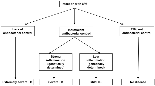
Background: Infection with Mycobacterium tuberculosis (Mtb) results in different clinical outcomes ranging from asymptomatic containment to rapidly progressing tuberculosis (TB). The mechanisms controlling TB progression in immunologically-competent hosts remain unclear.

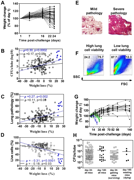
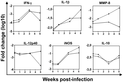
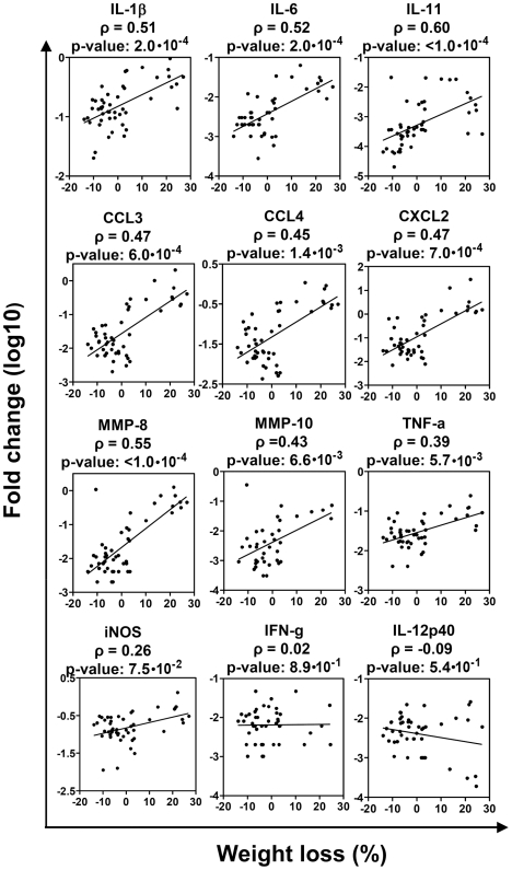
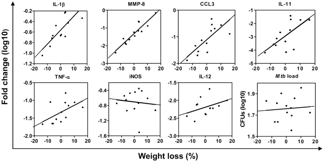
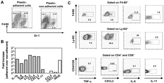
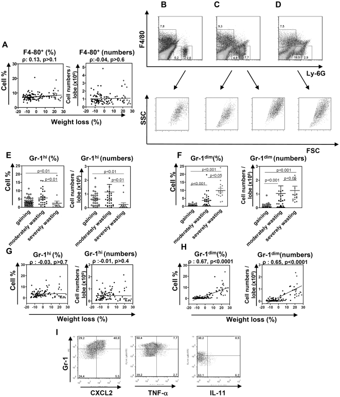
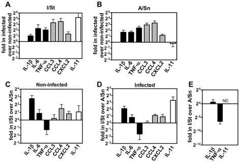
Methodology/principal findings: To address these mechanisms, we analyzed TB progression in a panel of genetically heterogeneous (A/SnxI/St) F2 mice, originating from TB-highly-susceptible I/St and more resistant A/Sn mice. In F2 mice the rates of TB progression differed. In mice that did not reach terminal stage of infection, TB progression did not correlate with lung Mtb loads. Nor was TB progression correlated with lung expression of factors involved in antibacterial immunity, such as iNOS, IFN-gamma, or IL-12p40. The major characteristics of progressing TB was high lung expression of the inflammation-related factors IL-1beta, IL-6, IL-11 (p<0.0003); CCL3, CCL4, CXCL2 (p<0.002); MMP-8 (p<0.0001). The major predictors of TB progression were high expressions of IL-1beta and IL-11. TNF-alpha had both protective and harmful effects. Factors associated with TB progression were expressed mainly by macrophages (F4-80(+) cells) and granulocytes (Gr-1(hi)/Ly-6G(hi) cells). Macrophages and granulocytes from I/St and A/Sn parental strains exhibited intrinsic differences in the expression of inflammatory factors, suggesting that genetically determined peculiarities of phagocytes transcriptional response could account for the peculiarities of gene expression in the infected lungs. Another characteristic feature of progressing TB was the accumulation in the infected lungs of Gr-1(dim) cells that could contribute to TB progression.

Conclusions/significance: In a population of immunocompetent hosts, the outcome of TB depends on quantitatively- and genetically-controlled differences in the intensity of inflammatory responses, rather than being a direct consequence of mycobacterial colonization. Local accumulation of Gr-1(dim) cells is a newly identified feature of progressing TB. High expression of IL-1beta and IL-11 are potential risk factors for TB progression and possible targets for TB immunomodulation.


Conflict of interest statement

Competing Interests: The authors have declared that no competing interests exist.

Figures

Figure 1
Figure 1. F2 mice display different rates of TB progression.
F2 mice were challenged i.t. with 103 CFU of Mtb. Weight was monitored once a week. A, Kinetics of weight change. 100% - weight on day 1 post-infection. Shown are representative results obtained in two (n = 52) independent experiments that included mice of both sexes. B–D, Mycobacterial load (B), lung pathology (C), and lung cell viability (D) in mice displaying different degree of wasting. Severely wasting mice (wasting by more than 20%) are indicated by blue circles. Lines show the predictions of the linear regression when all mice are included in the analysis (dashed blue lines) or when severely wasting mice are excluded from the analysis (solid black lines). ρ, Spearman correlation coefficient; p, p-value for ANOVA. E, F, Examples of lung tissue sections and lung cell flow cytometry representing two extremes with mild and severe pathology. Numbers in F indicate the percentages of dead (left) and live (right) cells. G, H, Long-term monitoring of F2 mice (n = 30, two independent experiments). G, The kinetics of weight change. H, Comparison of mycobacterial loads observed on days 24 and 140 post-infection.
Figure 2
Figure 2. TB-susceptible I/St and TB-resistant A/Sn mice infected with Mtb differ by the expression of genes associated with T-cell mediated response and inflammation.
I/St and A/Sn mice were challenged with Mtb or left un-infected. The expression of genes was analyzed in the lungs at weeks 0, 1, 3, and 5 post-challenge. Shown are typical examples of the expression of genes which were down-regulated in I/St lungs at late time-points post-challenge (IFN-γ, IL-12p40), up-regulated in I/St lungs at late time-points post-challenge (IL-1β, iNOS), up-regulated in I/St lungs at all time-points post-infection (MMP-8), and genes that were expressed similarly in I/St and A/Sn lungs (IL-10). Mean ±SD are shown (n = 3–4 per time-point). I/St, dashed line; A/Sn, solid line.
Figure 3
Figure 3. In F2 mice, TB progression correlates directly with lung expression of pro-inflammatory cytokines, chemokines, and metalloproteinases.
F2 mice were challenged with Mtb as described in Figure 1. On day 24, lung mRNA was extracted and assayed by real-time PCR. Shown are the correlations between wasting and lung mRNA expression of indicated genes in F2 female mice (n = 48).
Figure 4
Figure 4. F2 mice exhibiting similar mycobacterial loads display different rates of TB progression that positively correlate with the lung inflammatory response.
F2 mice challenged with Mtb were divided into several groups, each having similar Mtb loads. Correlations between wasting, mycobacterial load, and cytokine/chemokine expression in mice with lung mycobacterial burdens ranging from 3.3×10⁷ to 1×10⁸ CFUs/lobe are shown (for Spearman correlation coefficients and p-values see Table 3).
Figure 5
Figure 5. Pro-inflammatory factors associated with TB progression are expressed by lung phagocytic cells.
F2 mice were challenged with Mtb. On day 24 post-infection, suspensions of lung cells isolated from moderately wasting mice were separated into plastic-adherent and non-adherent populations, and gene expression was analyzed by real-time PCR. A, Flow cytometry analysis of plastic-adherent and plastic-non-adherent populations. Cells were stained with mAbs specific to F4-80 and Gr-1 (clone RB6-8C5) antigens. Note enrichment for F4/80+ and Gr-1+ cells in plastic-adherent over non-adherent population (60% over 14%). B, Gene expression in plastic-adherent versus plastic-nonadherent populations (fold change in expression). Closed bars: pro-inflammatory factors associated with TB progression; open bars: factors that did not correlate with TB progression (Figure 3, Table 2 and data not shown). C, Production of TNF-α, CXCL2, IL-6, and IL-11 by different populations of lung cells (intracellular staining, two independent experiments). Cells were stained with mAbs specific to CD4, CD8, F4-80, and Ly-6G (clone 1A8) antigens. Gates are placed based on the fluorescence-minus-one control for each of analyzed subset.
Figure 6
Figure 6. The accumulation of Gr-1dim cells in the infected lungs is a characteristic feature of progressive TB.
F2 mice were challenged with Mtb as described in Figure 1. On day 24, lung cell suspensions were obtained and stained with mAbs specific to F4-80 and Gr-1 (clone RB8-C6) or Ly-6G (clone 1A8) antigens (A–H). A, correlation between wasting, percentages and numbers of F4-80+Gr-1 cells in the lungs. B–D, typical examples of flow cytometry analysis of cells derived from the lungs of gaining (B) or wasting (C, D) mice. Shown are results obtained with PE-anti-F4-80 and FITC-anti-Ly-6G Abs. E, F, The content of Gr-1hi (E) and Gr-1dim (F) cells in the lungs of gaining, moderately wasting and severely wasting mice. G, H, correlation between wasting, percentages and numbers of Gr-1hi (G) and Gr-1dim (H) cells in the lungs. Note that Gr-1dim cells are negligible in gaining mice and that all mice with increased percentages or numbers of Gr-1dim cells are wasting. I, Intracellular cytokines in Gr-1hi and Gr-1dim cells. Note that CXCL2 and TNF-α are produced mainly by Gr-1hi cells, while IL-11 – by Gr-1dim cells.
Figure 7
Figure 7. I/St and A/Sn macrophages and neutrophils differ in the expression of pro-inflammatory cytokines.
A–D, Analysis of gene expression in macrophages. Macrophages were obtained from the peritoneal cavity of I/St and A/Sn mice, cultured with or without Mtb, and used for gene expression analysis. A, B, Gene expression in infected versus uninfected macrophages from I/St (A) and A/Sn (B) mice. C, D, Gene expression in I/St versus A/Sn macrophages, either uninfected (C) or infected (D). Data from one of three similar experiments are shown. E, Analysis of gene expression in neutrophils. Neutrophils were obtained from the peritoneal cavity of I/St and A/Sn mice and used for gene expression. Shown are gene expressions in I/St versus A/Sn neutrophils (results of three independent experiments). Bars show the relative expression of corresponding genes in infected versus un-infected phagocytes (A, B) or in I/St versus A/Sn phagocytes (C–E).
Figure 8
Figure 8. Suggested roles for antibacterial and inflammatory responses in the determination of TB outcome.
In hosts with deficiency in antibacterial immune response, progressive Mtb growth induces extremely severe TB. In hosts able to eradicate Mtb, no disease is developed. In hosts who are able to restrict Mtb growth but fail to completely eradicate the infection, the outcome depends on quantitatively- and genetically-determined peculiarities of inflammatory response.

Similar articles

Cited by

References

    1. Raviglione MC. The TB epidemic from 1992 to 2002. Tuberculosis (Edinb) 2003;83:4–14. - PubMed
    1. Bellamy R, Hill AVS. Host genetic susceptibility to human tuberculosis. Novartis Foundation Symposium. 1998;217:3–23. - PubMed
    1. Dietrich WF. Using mouse genetics to understand infectious disease pathogenesis. Genome Res. 2001;11:325–331. - PubMed
    1. Flynn JL, Ernst JD. Immune responses in tuberculosis. Curr Opin Immunol. 2000;12:432–436. - PubMed
    1. Collins HL, Kaufmann SH. The many faces of host responses to tuberculosis. Immunology. 2001;103:1–9. - PMC - PubMed

Publication types

MeSH terms