. 2010 May 3;5(5):e10467.
doi: 10.1371/journal.pone.0010467.

Role of PAF receptor in proinflammatory cytokine expression in the dorsal root ganglion and tactile allodynia in a rodent model of neuropathic pain

Affiliations

Role of PAF receptor in proinflammatory cytokine expression in the dorsal root ganglion and tactile allodynia in a rodent model of neuropathic pain

Shigeo Hasegawa et al. PLoS One. .

Abstract

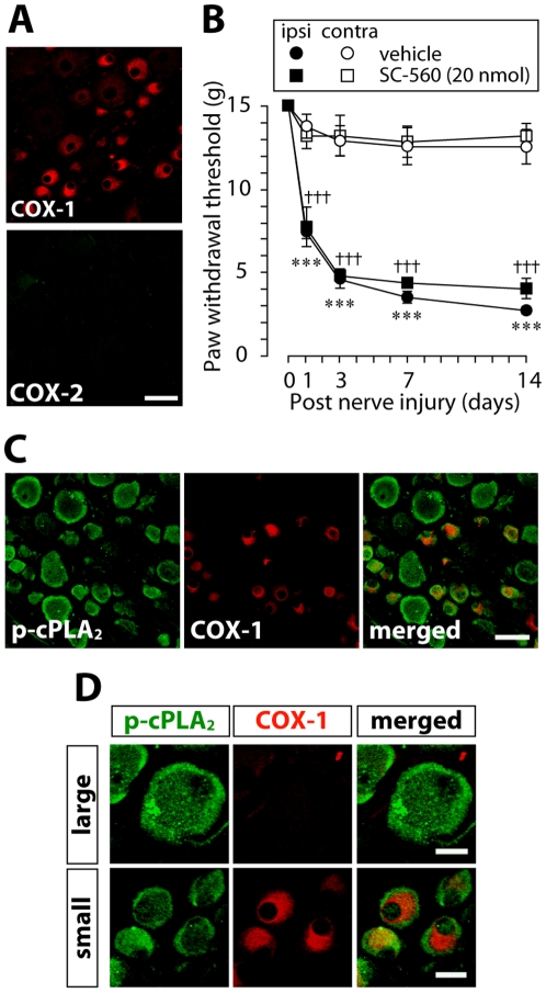
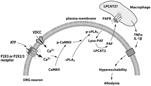
Background: Neuropathic pain is a highly debilitating chronic pain following damage to peripheral sensory neurons and is often resistant to all treatments currently available, including opioids. We have previously shown that peripheral nerve injury induces activation of cytosolic phospholipase A(2) (cPLA(2)) in injured dorsal root ganglion (DRG) neurons that contribute to tactile allodynia, a hallmark of neuropathic pain. However, lipid mediators downstream of cPLA(2) activation to produce tactile allodynia remain to be determined.

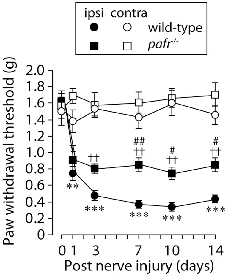
Principal findings: Here we provide evidence that platelet-activating factor (PAF) is a potential candidate. Pharmacological blockade of PAF receptors (PAFRs) reduced the development and expression of tactile allodynia following nerve injury. The expression of PAFR mRNA was increased in the DRG ipsilateral to nerve injury, which was seen mainly in macrophages. Furthermore, mice lacking PAFRs showed a reduction of nerve injury-induced tactile allodynia and, interestingly, a marked suppression of upregulation of tumor necrosis factor alpha (TNFalpha) and interleukin-1beta (IL-1beta) expression in the injured DRG, crucial proinflammatory cytokines involved in pain hypersensitivity. Conversely, a single injection of PAF near the DRG of naïve rats caused a decrease in the paw withdrawal threshold to mechanical stimulation in a dose-dependent manner and an increase in the expression of mRNAs for TNFalpha and IL-1beta, both of which were inhibited by pretreatment with a PAFR antagonist.

Conclusions: Our results indicate that the PAF/PAFR system has an important role in production of TNFalpha and IL-1beta in the DRG and tactile allodynia following peripheral nerve injury and suggest that blocking PAFRs may be a viable therapeutic strategy for treating neuropathic pain.


Conflict of interest statement

Competing Interests: The authors have declared that no competing interests exist.

Figures

Figure 1
Figure 1. A COX inhibitor does not prevent the development of tactile allodynia after nerve injury.
A. Immunofluorescence labeling of COX-1 and COX-2 proteins in the L5 DRG 14 days after nerve injury demonstrated that immunoreactivity of COX-1, but not COX-2, was present mainly in small-sized neurons. Scale bar, 50 µm. B. The COX-1 inhibitor SC-560 (20 nmol/10 µl) administered through a catheter whose tip was positioned near the L5 DRG once daily for 14 days did not suppress the development of nerve injury-induced a decrease in the paw withdrawal threshold of tactile stimulation using von Frey filaments. ***p<0.001 compared with the threshold of the vehicle-treated group on day 0. †††p<0.001 compared with the threshold of the inhibitor-treated group on day 0. Data are presented as mean ± SEM of the paw withdrawal threshold of five animals. C. Double immunofluorescence labeling of p-cPLA2 with COX-1 in L5 DRG neurons 14 days after nerve injury showed that most of neurons with the translocated p-cPLA2 were not double-labeled with COX-1-IR. Scale bar, 50 µm. D. Highly magnified pictures of large and small diameter DRG neurons demonstrated that in large neurons, p-cPLA2 was not co-expressed with COX-1-IR. In small neurons, the subcellular localizations of p-cPLA2 and COX-1-IR were different: COX-1-immunoreactivity was observed in the perinuclear area where p-cPLA2- immunoreactivity was not accumulated. Scale bars, 20 µm.
Figure 2
Figure 2. LOX inhibitors do not prevent the development of tactile allodynia after nerve injury.
A, B. The 5-LOX inhibitor AA-861 (20 nmol/10 µl) (A) and the 12- and 15-LOX inhibitor baicalein (20 nmol/10 µl) (B) administered through a catheter whose tip was positioned near the L5 DRG once daily for 14 days did not suppressed the development of nerve injury-induced tactile allodynia. **p<0.01, ***p<0.001 compared with the threshold of the vehicle-treated group on day 0. ††p<0.01, †††p<0.001 compared with the threshold of the inhibitor-treated group on day 0. All data are presented as mean ± SEM of the paw withdrawal threshold of five animals.
Figure 3
Figure 3. PAFR and LPAR antagonists suppress the development of tactile allodynia by injury to the L5 spinal nerve.
A, B. Administration of PAFR antagonist CV-3988 (10 nmol/10 µl) (A) and the LPAR antagonist Ki16425 (10 nmol/10 µl) (B) through a catheter whose tip was positioned near the L5 DRG once daily for 14 days after nerve injury suppressed the development of tactile allodynia. **p<0.01, ***p<0.001 compared with the threshold of the vehicle-treated group on day 0. ††p<0.01, †††p<0.001 compared with the threshold of the antagonist-treated group on day 0. #p<0.05, ##p<0.01, ###p<0.001 compared with the threshold of the vehicle-treated group at each time point. All data are presented as mean ± SEM of the paw withdrawal threshold of five to eight animals.
Figure 4
Figure 4. Inhibition of PAFR induces a relieving effect on existing tactile allodynia.
A. The decrease in paw withdrawal threshold was attenuated by a single administration of CV-3988 (10 nmol/10 µl) (A), but not Ki16425 (10 nmol/10 µl) (B), through a catheter whose tip was positioned near the L5 DRG on day 7 after nerve injury. ***p<0.001, †††p<0.001 compared with pre-injury baseline (Pre). ###p<0.001 compared with the threshold on day 7. n.s. means “not significant”. All data are presented as mean ± SEM of the paw withdrawal threshold of five to eight animals.
Figure 5
Figure 5. LPCAT2 is expressed in DRG neurons positive to p-cPAL2 following peripheral nerve injury.
Immunohistochemical analysis using two adjacent DRG sections (singly immunostained one section with p-cPLA2 or LPCAT2 antibody) revealed that there were DRG neurons positive for both p-cPLA2 and LPCAT2 in the injured DRG (arrowheads) 7 days after nerve injury. Arrows indicate unknown cells that were strongly positive to LPCAT2-IR and negative to p-cPLA2.-IR. Scale bar, 50 µm.
Figure 6
Figure 6. PAFR mRNA is upregulated in the DRG following peripheral nerve injury.
A. Real-time PCR analysis revealed that PAFR mRNA expression in total RNA extract from the L5 DRG was markedly increased after peripheral nerve injury. The bar graphs show the average fold increase in the level of PAFR mRNA expression in the DRG compared with the mean expression level of PAFR mRNA in naïve rats. Each measurement was normalized to GAPDH content. **p<0.01, ***p<0.001 compared with naïve rats. ††p<0.01, †††p<0.001 compared with the contralateral side. All data are presented as mean ± SEM of five individual animals. B-D. DIG-labeled antisense (B: contralateral, C: ipsilateral) and sense (D: ipsilateral) probes specific for PAFR mRNA were visualized by in situ hybridization in the rat DRG 7 days after nerve injury. Strong PAFR mRNA signals were observed in the cells surrounding DRG neurons in the ipsilateral DRG 7 days after nerve injury. Arrowheads show PAFR mRNA-positive cells. ‘N’ indicates neuronal cells. Similar results were observed in each of three experiments. Scale bar, 50 µm. E,F. In situ hybridization combined with immunohistochemistry for the macrophages/microglia marker Iba1 and the satellite glia marker GFAP was performed. PAFR mRNA signals overlapped with Iba1-IR (arrowheads, E) but not with GFAP-IR (GFAP: white arrowheads, PAFR: white arrows, F). Scale bar, 25 µm.
Figure 7
Figure 7. Deletion of pafr reduces tactile allodynia.
While wild-type mice with an L5 nerve injury showed a progressively decreased paw withdrawal threshold, pafr −/− mice showed an attenuation of the decrease in paw withdrawal threshold after nerve injury. **p<0.01, ***p<0.001 compared with the threshold of wild-type mice on day 0. ††p<0.01 compared with the threshold of pafr −/− mice on day 0. #p<0.05, ##p<0.01 compared with the threshold of the wild-type mice at each time point. All data are presented as mean ± SEM of six to eight animals.
Figure 8
Figure 8. Upregulation of TNFα and IL-1β gene expression in the DRG is reduced in pafr −/− mice.
A, B. Real-time PCR analysis demonstrated that following nerve injury, the expression of TNFα (A) and IL-1β (B) mRNAs in the ipsilateral DRG of wild-type mice was much higher than in the contralateral side on day 7. However, pafr −/− mice failed to show upregulation of these cytokines and there were significant differences in these expressions of the ipsilateral side between wild-type and pafr −/− mice. Each measurement was normalized to 18S mRNA content. **p<0.01, ***p<0.001 compared with the contralateral side of wild-type mice. #p<0.05, ##p<0.01 compared with the ipsilateral side. n.s. means “not significant”. All data are presented as mean ± SEM of six to eight animals.
Figure 9
Figure 9. Injection of PAF near the DRG induces a decrease in the paw withdrawal threshold and upregulation of TNFα and IL-1β mRNAs.
A. Paw withdrawal threshold was decreased by single administration of several different doses of PAF near the DRG in normal rats. *p<0.05, **p<0.01, ***p<0.001 compared with the threshold of the vehicle-treated group. B. The PAF-induced decrease in paw withdrawal threshold was suppressed by pretreatment with CV-3988. ***p<0.001 compared with the threshold of the vehicle-treated group. ##p<0.01 compared with the threshold of the PAF-treated group. n.s. means “not significant”. C, D. Real-time PCR analysis showed that administration of PAF near the DRG increased expression of TNFα (C) and IL-1β (D) mRNAs in the DRG 45 min after the administration. The bar graphs show the average fold increase in the level of TNFα and IL-1β mRNAs in the DRG compared with the mean expression level of these mRNAs in the vehicle-treated group. Each measurement was normalized to 18S mRNA content. *p<0.05, **p<0.01, ***p<0.001 compared with the vehicle-treated group. #p<0.05, ###p<0.001 compared with the PAF-treated group. n.s. means “not significant”. All data are presented as mean ± SEM of four to six animals.
Figure 10
Figure 10. Schematic representation of the proposed mechanism underlying the PAF/PAFR system-mediated neuropathic pain after peripheral nerve injury.
After peripheral nerve injury, cPLA2 is activated by Ca2+ signaling evoked by P2X3 or P2X2/3 receptors (subtype of ionotropic purinergic receptors) and voltage-gated Ca2+ channels (VDCC). The Ca2+-dependent cPLA2 activation involves Ca2+/calmodulin-dependent protein kinase II (CaMKII). cPLA2 supply lyso-PAF which in turn converts into PAF by the lyso-PAF-acetyltransferase LPCAT2. PAF activates PAFR expressed in macrophages. Activation of PAFR may lead to production and release of proinflammatory cytokines, TNFα and IL-1β. These cytokines may increase the excitability of DRG neurons that link to nerve injury-induced tactile allodynia.

References

    1. Woolf CJ, Mannion RJ. Neuropathic pain: aetiology, symptoms, mechanisms, and management. Lancet. 1999;353:1959–1964. - PubMed
    1. Woolf CJ, Salter MW. Neuronal plasticity: increasing the gain in pain. Science. 2000;288:1765–1769. - PubMed
    1. Scholz J, Woolf CJ. Can we conquer pain? Nat Neurosci. 2002;5(Suppl):1062–1067. - PubMed
    1. Shimizu T, Ohto T, Kita Y. Cytosolic phospholipase A2: biochemical properties and physiological roles. IUBMB Life. 2006;58:328–333. - PubMed
    1. Tsuda M, Hasegawa S, Inoue K. P2X receptors-mediated cytosolic phospholipase A2 activation in primary afferent sensory neurons contributes to neuropathic pain. J Neurochem. 2007;103:1408–1416. - PubMed

Publication types

MeSH terms

Substances