. 2010:2010:940383.
doi: 10.1155/2010/940383. Epub 2010 May 3.

Normal human gingival epithelial cells sense C. parapsilosis by toll-like receptors and module its pathogenesis through antimicrobial peptides and proinflammatory cytokines

Affiliations

Normal human gingival epithelial cells sense C. parapsilosis by toll-like receptors and module its pathogenesis through antimicrobial peptides and proinflammatory cytokines

Raouf Bahri et al. Mediators Inflamm. 2010.

Abstract

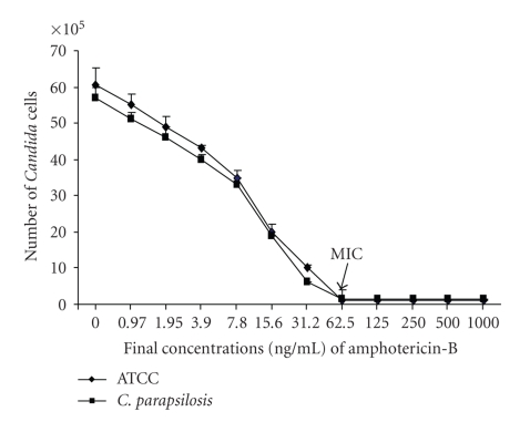
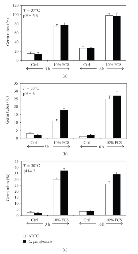
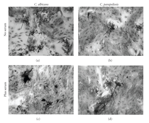
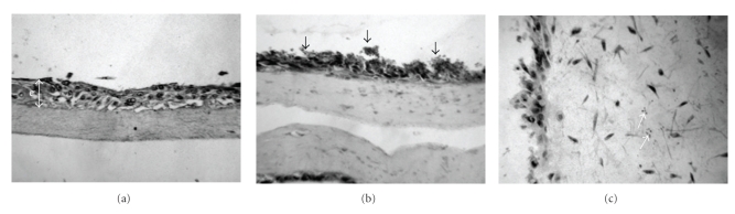
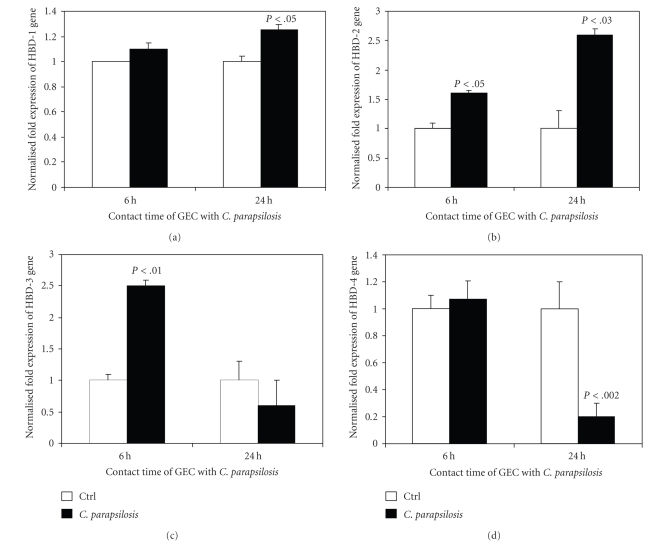
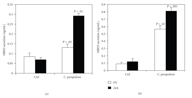
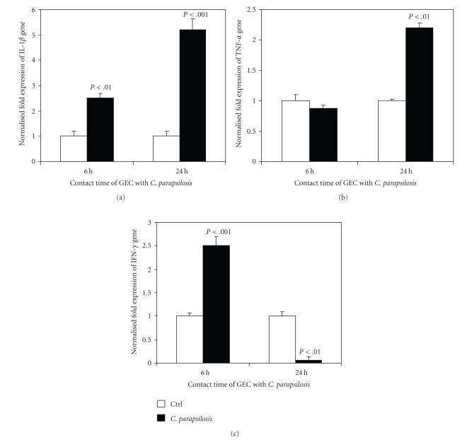
This study was designed to investigate the interaction between C. parapsilosis and human epithelial cells using monolayer cultures and an engineered human oral mucosa (EHOM). C. parapsilosis was able to adhere to gingival epithelial cells and to adopt the hyphal form in the presence of serum. Interestingly, when cultured onto the engineered human oral mucosa (EHOM), C. parapsilosis formed small biofilm and invaded the connective tissue. Following contact with C. parapsilosis, normal human gingival epithelial cells expressed high levels of Toll-like receptors (TLR)-2, -4, and -6, but not TLR-9 mRNA. The upregulation of TLRs was paralleled by an increase of IL-1beta, TNFalpha, and IFNgamma mRNA expression, suggesting the involvement of these cytokines in the defense against infection with C. parapsilosis. The active role of epithelial cells in the innate immunity against C. parapsilosis infection was enhanced by their capacity to express high levels of human beta-defensin-1, -2, and -3. The upregulation of proinflammatory cytokines and antimicrobial peptide expression may explain the growth inhibition of C. parapsilosis by the gingival epithelial cells. Overall results provide additional evidence of the involvement of epithelial cells in the innate immunity against C. parapsilosis infections.


Figures

Figure 1
Figure 1
Sensitivity of C. parapsilosis to amphotericin-B. The yeast was cultured in the presence of various concentrations of amphotericin-B for 24 hours and its growth was assessed by MTT assay. Quantification of live C. parapsilosis was obtained using a standard live curve. The effect of the antifungal agent on C. parapsilosis was compared to that on C. albicans. Results are presented as means ± SD of six different experiments.
Figure 2
Figure 2
Blastospore-to-hyphal transition of C. parapsilosis. Following culture at different temperatures and neutral pH with or without proteins (serum), the percentage of C. parapsilosis germ tubes was determined by dividing the number of hyphae over the total number of cells (blastospore and hyphae) in each culture condition. The mean relative values for six separate experiments are shown. C. albicans was used as a reference strain to C. parapsilosis.
Figure 3
Figure 3
C. parapsilosis adhesion to the gingival epithelial monolayer culture. Human gingival cells were grown up to 80% confluence, pulsed with C. parapsilosis or C. albicans, and incubated for 6 hours with or without serum. Following this culture period, the supernatant containing nonattached Candida was discarded, and the cultures were washed and stained with Cristal violet. Representative photographs are presented. Magnification: 250×.
Figure 4
Figure 4
Effect of C. parapsilosis on tissue structure and the formation of a biofilm on the engineered human oral mucosa. Histological features of EHOM following exposure to C. parapsilosis. EHOM tissue was infected with (a) no Candida cells (uninfected control) and (b, c) C. parapsilosis strain. Note the formation of a biofilm on the tissue (b) and the presence of C. parapsilosis in the connective tissue (c). Representative photographs of four different experiments are shown (two EHOMs per experiment). Magnification ×250 for parts (a) and (b), and ×600 for part (c). e: epithelium.
Figure 5
Figure 5
Quantification of mRNA expression levels of TLR-2, -4, -6, and -9 by gingival epithelial cells following infection with C. parapsilosis. Gingival epithelial cells were cultured with or without C. parapsilosis for 6 and 24 hours. Total RNA content was extracted from the cells following each culture period and was used for quantitative RT-PCR of the TLR-2, -4, -6, and -9 genes, as described in Section 2. Results are presented as a fold expression of the gene in the test sample compared to this gene expression in the control. Data are expressed as means ± SD from triplicate assays of three different experiments. (a) TLR-2 expression, (b) TLR-4 expression, (c) TLR-6 expression, and (d) TLR-9 expression. GEC: gingival epithelial cells.
Figure 6
Figure 6
Quantification of mRNA expression levels of HBD-1, -2, -3, and -4 by gingival epithelial cells following infection with C. parapsilosis. Gingival epithelial cells were cultured with or without C. parapsilosis for 6 and 24 hours. Total RNA was extracted from the cells following each culture period and was used for quantitative RT-PCR of the HBD-1, -2, -3, and -4 genes, as described in Section 2. Results are presented as a fold expression of the gene in the test sample compared to this gene expression in the control. Data are expressed as means ± SD from triplicate assays of three different experiments. (a) HBD-1 expression, (b) HBD-2 expression, (c) HBD-3 expression, and (d) HBD-4 expression. GEC: gingival epithelial cells.
Figure 7
Figure 7
Quantification of HBD2 and HBD3 levels secreted by gingival epithelial cells following infection with C. parapsilosis. Gingival epithelial cells were cultured with or without C. parapsilosis for 6 and 24 hours. Supernatants were collected and used to quantify HBD2 (a) and HBD3 (b) using ELISA kits. Data are means ± SD of three separate experiments. The levels of significance were obtained by comparing the amount of AMP obtained with unstimulated samples to that obtained with samples stimulated C. parapsilosis.
Figure 8
Figure 8
Quantification of mRNA expression levels of IL-1β, TNFα, and IFNγ by gingival epithelial cells following infection with C. parapsilosis. Gingival epithelial cells were cultured with or without C. parapsilosis for 6 and 24 hours. Total RNA was extracted from the cells following each culture period and was used for quantitative RT-PCR of the IL-1β, TNFα, and IFNγ genes, as described in Section 2. Results are presented as a fold expression of the gene in the test sample compared to this gene expression in the control. Data are expressed as means ± SD from triplicate assays of three different experiments. (a) IL-1β expression, (b) TNFα expression, and (c) IFNγ expression. GEC: gingival epithelial cells.

References

    1. Lupetti A, Danesi R, van’t Wout JW, van Dissel JT, Senesi S, Nibbering PH. Antimicrobial peptides: therapeutic potential for the treatment of candida infections. Expert Opinion on Investigational Drugs. 2002;11(2):309–318. - PubMed
    1. Sobel JD. The emergence of non-albicans Candida species as causes of invasive candidiasis and candidemia. Current Infectious Disease Reports. 2006;8(6):427–433. - PubMed
    1. Weems JJ., Jr. Candida parapsilosis: epidemiology, pathogenicity, clinical manifestations, and antimicrobial susceptibility. Clinical Infectious Diseases. 1992;14(3):756–766. - PubMed
    1. Fell JW, Meyer SA. Systematics of yeast species in the Candida parapsilosis group. Mycopathologia et Mycologia Applicata. 1967;32(3):177–193. - PubMed
    1. Clark TA, Slavinski SA, Morgan J, et al. Epidemiologic and molecular characterization of an outbreak of Candida parapsilosis bloodstream infections in a community hospital. Journal of Clinical Microbiology. 2004;42(10):4468–4472. - PMC - PubMed

Publication types

MeSH terms