. 2010 May 3;5(5):e10436.
doi: 10.1371/journal.pone.0010436.

Agonist anti-GITR monoclonal antibody induces melanoma tumor immunity in mice by altering regulatory T cell stability and intra-tumor accumulation

Affiliations

Agonist anti-GITR monoclonal antibody induces melanoma tumor immunity in mice by altering regulatory T cell stability and intra-tumor accumulation

Adam D Cohen et al. PLoS One. .

Abstract

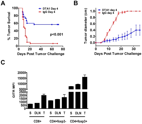
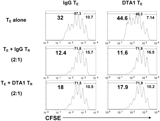
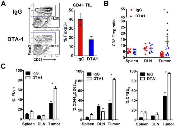
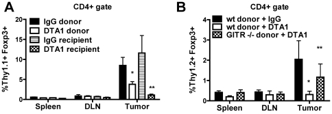
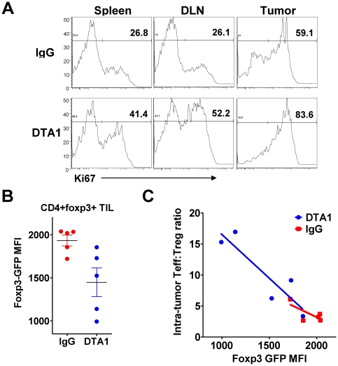
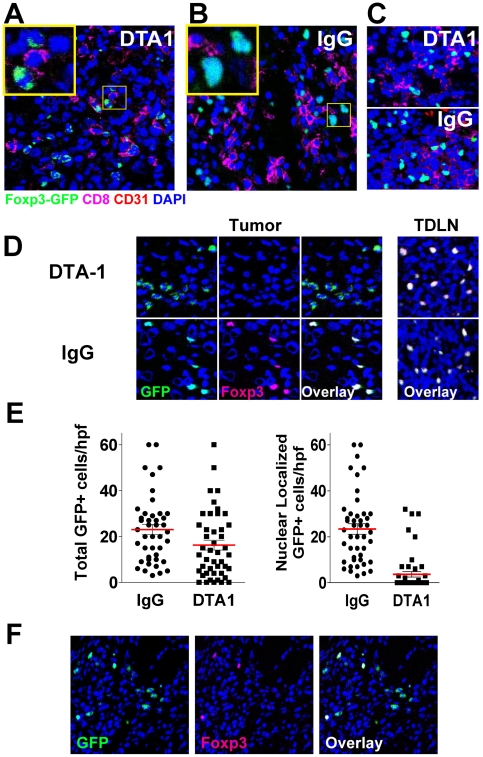
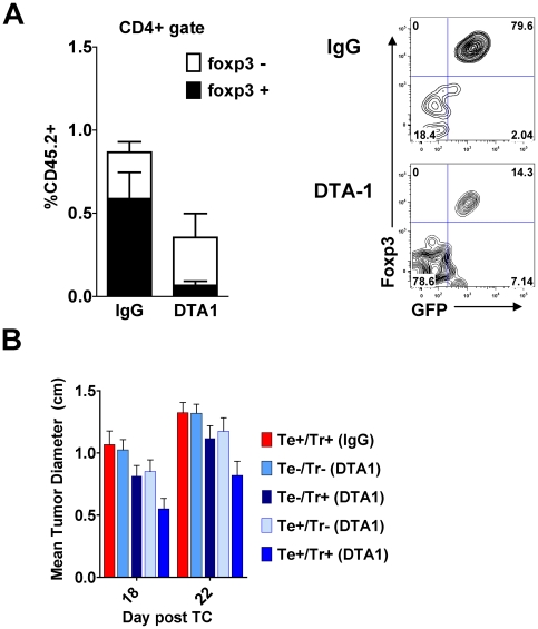
In vivo GITR ligation has previously been shown to augment T-cell-mediated anti-tumor immunity, yet the underlying mechanisms of this activity, particularly its in vivo effects on CD4+ foxp3+ regulatory T cells (Tregs), have not been fully elucidated. In order to translate this immunotherapeutic approach to the clinic it is important gain better understanding of its mechanism(s) of action. Utilizing the agonist anti-GITR monoclonal antibody DTA-1, we found that in vivo GITR ligation modulates regulatory T cells (Tregs) directly during induction of melanoma tumor immunity. As a monotherapy, DTA-1 induced regression of small established B16 melanoma tumors. Although DTA-1 did not alter systemic Treg frequencies nor abrogate the intrinsic suppressive activity of Tregs within the tumor-draining lymph node, intra-tumor Treg accumulation was significantly impaired. This resulted in a greater Teff:Treg ratio and enhanced tumor-specific CD8+ T-cell activity. The decreased intra-tumor Treg accumulation was due both to impaired infiltration, coupled with DTA-1-induced loss of foxp3 expression in intra-tumor Tregs. Histological analysis of B16 tumors grown in Foxp3-GFP mice showed that the majority of GFP+ cells had lost Foxp3 expression. These "unstable" Tregs were absent in IgG-treated tumors and in DTA-1 treated TDLN, demonstrating a tumor-specific effect. Impairment of Treg infiltration was lost if Tregs were GITR(-/-), and the protective effects of DTA-1 were reduced in reconstituted RAG1(-/-) mice if either the Treg or Teff subset were GITR-negative and absent if both were negative. Our results demonstrate that DTA-1 modulates both Teffs and Tregs during effective tumor treatment. The data suggest that DTA-1 prevents intra-tumor Treg accumulation by altering their stability, and as a result of the loss of foxp3 expression, may modify their intra-tumor suppressive capacity. These findings provide further support for the continued development of agonist anti-GITR mAbs as an immunotherapeutic strategy for cancer.


Conflict of interest statement

Competing Interests: The authors have declared that no competing interests exist.

Figures

Figure 1
Figure 1. Upregulation of GITR expression correlates with optimal timing of single dose DTA-1 therapy.
A and B. C57BL/6 mice (n = 10/group) were challenged intradermally with 50,000 B16 melanoma cells and treated with 1 mg DTA-1 or rat IgG i.p. on day 4 after tumor challenge. Tumor survival (A) and mean tumor diameter + SEM over time is depicted (B) C. Untreated mice (n = 3/group) bearing 4 day-old B16 matrigel tumors (500,000 cells) were sacrificed and lymphocytes isolated from spleens (S), tumor-draining lymph nodes (DLN), and tumors (T), were stained for CD4, CD8, foxp3, and GITR. Mean GITR fluorescence intensity (MFI) and SEM within each T cell subset is depicted.
Figure 2
Figure 2. In vivo GITR ligation does not globally alter capacity of Tregs to suppress or CD8+ Teffs to resist suppression.
B16-bearing C57BL/6 mice (n = 6/group) received DTA-1 or IgG on day 4. TDLN were harvested on day 7 and CD8+ (Teff) and CD4+CD25+ (Treg) cells were isolated by MACS beads. CFSE-labeled, IgG- or DTA-1-treated Teff were cultured with IgG- or DTA-1-treated Treg at 2∶1 Teff:Treg ratio, with irradiated APCs and anti-CD3 mAb 1 µg/ml for 4 days. A. Representative sample showing CFSE dilution in CD8+DAPI- cells.
Figure 3
Figure 3. GITR ligation by DTA-1 limits Treg accumulation within the tumor and enhances intra-tumor CD8+ T-cell activity.
A. and B. B16-bearing mice treated with DTA-1 or IgG on day 4 had spleens, TDLN, and tumors harvested on day 10 and lymphocytes analyzed by FACS. A. Representative FACS plots with gate frequencies, (left) and mean +SEM for frequency of Tregs within live CD4+ TIL gate (right) are shown. B. Ratio of CD8+ to CD4+foxp3+ cells in spleen, TDLN, and tumor. *p = 0.05 compared with IgG tumor. Pooled data from 3 independent experiments are shown. C. Naïve C57BL/6 mice (n = 3−5/group) received 4×10⁶ CFSE-labeled pmel-1 Thy1.1+CD8+ T cells 1 day prior to B16 inoculation. Recipients received DTA-1 or IgG on day 4, and donor pmel-1 CD8+ cells analyzed in spleens, TDLN, and tumors on day 14. Mean frequency +SEM is shown for IFNγ+ (left), activated CD44hiCD62Llo phenotype (center) and proliferation by CFSE dilution (right) of transferred pmel-1 T cells is shown. For IFNγ recall assay (C, left), lymphocytes from spleen, TDLN, and tumor were re-stimulated for 6 hours with irradiated, gp10025-33 peptide-pulsed EL4 cells. Background IFNγ production for lymphocytes cultured with unpulsed EL4 cells was <1%. Over 85% of IFNγ+ cells were also CD107a+ (data not shown). * p<0.05 compared with IgG group. Representative of 3 independent experiments.
Figure 4
Figure 4. DTA-1 reduces Treg tumor trafficking in a cell intrinsic manner.
A. B16-bearing Thy1.1+ donor mice were treated on day 5 with DTA-1 (“DTA-1 donor”) or IgG (“IgG donor”), and Tregs isolated on day 8 from their spleens and TDLN were transferred (0.7×10⁶ per recipient) to B16-bearing Thy1.2+ mice, treated 1 day earlier with cyclophosphamide 250 mg/kg i.p. Some recipients received Tregs from untreated B16-bearing donors and were then treated with DTA-1 (“DTA-1 recipient”) or IgG (“IgG recipient”) 12 hours after adoptive transfer. Recipient spleens, TDLN, and tumors were harvested 4 days after transfer. The percentage + SEM of donor (Thy1.1+CD4+foxp3+) Tregs within the total live CD4+ gate is depicted. *p = 0.05 compared with IgG donor, **p = 0.04 compared with IgG recipient. B. Tregs from Day 8 B16-bearing Thy1.2+ GITR−/− (“KO donor”) or GITR+/+ (“wt donor”) donors were transferred into day 8 B16-bearing Thy1.1+ recipients treated 1 day earlier with cyclophosphamide 250 mg/kg i.p. Recipients received DTA-1 or IgG 12 hours post-transfer, and spleens, TDLN, and tumors were harvested 4 days later. The % + SEM of donor (Thy1.2+CD4+foxp3+) Tregs within the total live CD4+ gate is depicted. *p = 0.07 compared with wt donor + IgG, **p = 0.24 compared with wt donor + IgG.
Figure 5
Figure 5. Intra-tumor Tregs show increased proliferation and lower Foxp3GFP MFI after DTA-1 treatment.
A. Representative Ki67 expression within live CD4+foxp3+ cells isolated from day 10 B16-bearing C57BL/6 mice treated with DTA-1 or IgG on day 4. B. and C. TIL were isolated from day 10 B16-bearing foxp3 GFP mice (n = 5/group) treated with DTA-1 or IgG on day 4 and examined by FACS. B. Mean fluorescence intensity (MFI) of GFP on live intra-tumor CD4+GFP+ cells. C. Intra-tumor CD8:Treg ratio of DTA-1- and IgG treated mouse in (B) is plotted against its foxp3GFP MFI, demonstrating a significant inverse correlation between Teff:Treg ratio and foxp3GFP MFI in DTA-1 treated mice only (p = 0.03). Data are representative of 3 independent experiments.
Figure 6
Figure 6. DTA-1-treated mice have abnormal intra-tumor Tregs which have lost foxp3 expression.
Tumors were harvested from day 10 B16-bearing foxp3 GFP mice (n = 4/group) treated with DTA-1 or IgG on day 4. Tumor sections were stained with anti-CD8 (magenta), anti-CD31 (red, to visualize endothelium), and DAPI (blue, for nuclear staining) and analyzed by immunofluorescence. A. DTA-1-treated tumor showing Tregs with irregular borders, weaker foxp3/GFP+ signal, and non-nuclear GFP localization (inset). B. IgG-treated tumor showing Tregs with foxp3 protein (GFP+, green) co-localizing with nucleus. C. DTA-1- and IgG-treated TDLN demonstrating intact foxp3+ cells. D. Left, top: DTA1-treated tumor co-stained with anti-foxp3. Note lack of foxp3 co-staining in cells with abnormal cytoplasmic GFP signal, compared with overlapping nuclear foxp3 and GFP in IgG-treated intra-tumor Tregs (left, bottom) or DTA-1- or IgG-treated TDLN (right). Scale for all images is show in A (bar = 25 µm). E. The number of GFP+ cells per high-powered field (hpf) were counted, regardless of either intensity or localization (“total”, left graph) or only with bright nuclear GFP signal (i.e. co-localizing with DAPI) (“nuclear GFP,” right graph). Pooled data from a total of 46 (IgG) or 48 (DTA-1) examined hpf (10–12 hpf per tumor ×4 tumors/group) are shown. F. Although the majority of DTA-1 treated Tregs have an “abnormal” GFP+ foxp3- phenotype, “intact” and “abnormal” Tregs can be found together within a minority of DTA-1 treated tumor sections.
Figure 7
Figure 7. DTA-1-induces foxp3 loss in intra-tumor Tregs after transfer, and the effects of DTA-1 require GITR expression on effector and regulatory T cells.
A. CD4+GFP+ Tregs isolated from spleens and TDLN of B16-bearing CD45.2+ foxp3gfp donor mice on day 8 after tumor challenge were transferred (0.7×10⁶ per recipient) into B16-bearing CD45.1+ mice treated with cyclophosphamide and DTA-1 or IgG, as described in Figure 4. Tumor-infiltrating lymphocytes were isolated 2–3 days later and CD45.2+ donor T cells were assessed for their level of foxp3 and GFP expression by FACS. Left: The % + SEM of donor (CD45.2+) Tregs found within the total live CD4+ gate is depicted, along with relative proportions that became foxp3-negative. Donor Tregs within DTA-1-treated tumors showed a significantly greater reduction in foxp3 expression (p = 0.001) Right:. Representative FACS plots gated on live CD45.2+CD4+ TIL. Data are pooled (n = 7 per group) from 2 independent experiments. B RAG1−/− mice (n = 5−9/group) were reconstituted with indicated combinations of effector (Te: CD8+ and CD4+CD25-) and regulatory (Tr: CD4+CD25+) T cells from GITR−/− (−) or GITR+/+ (+) littermates (See Figure S5 for schema). After 4 weeks, mice were challenged with B16 and treated with DTA-1 or IgG on day 4. At 18 and 22 days post-challenge, when all tumor-free survival is lost in WT untreated animals (Figure 1A), mean diameter of the Te+/Tr+ (DTA1) group was significantly different from all other groups. (p<0.05; two-tailed student's t-test). Data are pooled from 2 independent experiments.

References

    1. Nocentini G, Riccardi C. GITR: a multifaceted regulator of immunity belonging to the tumor necrosis factor receptor superfamily. Eur J Immunol. 2005;35:1016–1022. - PubMed
    1. Kohm AP, Williams JS, Miller SD. Cutting edge: ligation of the glucocorticoid-induced TNF receptor enhances autoreactive CD4+ T cell activation and experimental autoimmune encephalomyelitis. J Immunol. 2004;172:4686–4690. - PubMed
    1. Kanamaru F, Youngnak P, Hashiguchi M, Nishioka T, Takahashi T, et al. Costimulation via glucocorticoid-induced TNF receptor in both conventional and CD25+ regulatory CD4+ T cells. J Immunol. 2004;172:7306–7314. - PubMed
    1. Ronchetti S, Zollo O, Bruscoli S, Agostini M, Bianchini R, et al. GITR, a member of the TNF receptor superfamily, is costimulatory to mouse T lymphocyte subpopulations. Eur J Immunol. 2004;34:613–622. - PubMed
    1. Tone M, Tone Y, Adams E, Yates SF, Frewin MR, et al. Mouse glucocorticoid-induced tumor necrosis factor receptor ligand is costimulatory for T cells. Proc Natl Acad Sci U S A. 2003;100:15059–15064. - PMC - PubMed

Publication types

MeSH terms

Substances