. 2010 May 3;5(5):e10462.
doi: 10.1371/journal.pone.0010462.

Proteomic profiling of human keratinocytes undergoing UVB-induced alternative differentiation reveals TRIpartite Motif Protein 29 as a survival factor

Affiliations

Proteomic profiling of human keratinocytes undergoing UVB-induced alternative differentiation reveals TRIpartite Motif Protein 29 as a survival factor

Véronique Bertrand-Vallery et al. PLoS One. .

Abstract

Background: Repeated exposures to UVB of human keratinocytes lacking functional p16(INK-4a) and able to differentiate induce an alternative state of differentiation rather than stress-induced premature senescence.

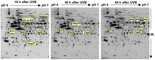
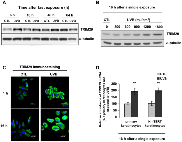
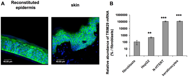
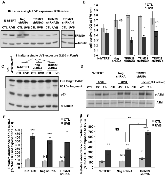
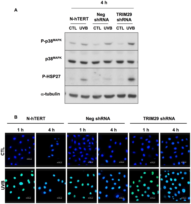
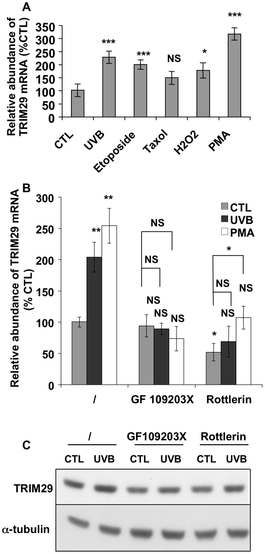
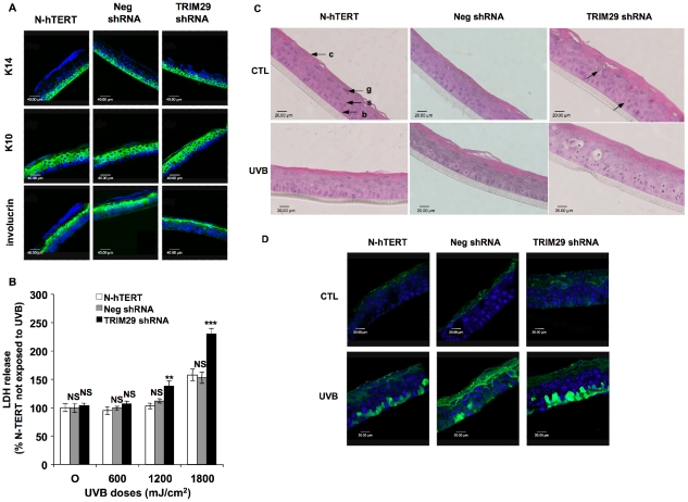
Methodology/principal findings: A 2D-DIGE proteomic profiling of this alternative state of differentiation was performed herein at various times after the exposures to UVB. Sixty-nine differentially abundant protein species were identified by mass spectrometry, many of which are involved in keratinocyte differentiation and survival. Among these protein species was TRIpartite Motif Protein 29 (TRIM29). Increased abundance of TRIM29 following UVB exposures was validated by Western blot using specific antibody and was also further analysed by immunochemistry and by RT-PCR. TRIM29 was found very abundant in keratinocytes and reconstructed epidermis. Knocking down the expression of TRIM29 by short-hairpin RNA interference decreased the viability of keratinocytes after UVB exposure. The abundance of involucrin mRNA, a marker of late differentiation, increased concomitantly. In TRIM29-knocked down reconstructed epidermis, the presence of picnotic cells revealed cell injury. Increased abundance of TRIM29 was also observed upon exposure to DNA damaging agents and PKC activation. The UVB-induced increase of TRIM29 abundance was dependent on a PKC signaling pathway, likely PKCdelta.

Conclusions/significance: These findings suggest that TRIM29 allows keratinocytes to enter a protective alternative differentiation process rather than die massively after stress.


Conflict of interest statement

Competing Interests: NB and MS are members of the StratiCELL SA. The authors confirm that this does not alter their adherence to all the PLoS ONE policies on sharing data and materials, as detailed online in the guide for authors.

Figures

Figure 1
Figure 1. Representative 2D-DIGE profiling of N-hTERT keratinocytes repeatedly exposed to UVB and remarkable differential spots intensities.
N-hTERT keratinocytes were exposed to UVB at 300 mJ/cm2 with 4 exposures per day for 2 days. Control samples were submitted to the same culture conditions without UVB. Three independent cultures (N = 3) were lysed at 16, 40 and 64 h after the 8th exposure to UVB. Labeled samples were mixed, set to isoelectrofocusing and second-dimension was performed. The CyDye images presented correspond to a typical 2D-DIGE protein pattern [cyanine fluorophore specific (Cy2) image] (M.W. molecular weight). Spots with significantly different intensity between control and UVB exposed cells (41, 34 and 30 variations at 16, 40 and 64 h respectively) are shown by circles drawn on spot boundaries (p<0.05, Student's t test in the three independent experiments). These spots were identified by nanoLC-MS-MS. Only a few proteins of remarkable interest are indicated here.
Figure 2
Figure 2. Increased abundance of TRIM29 after single or repeated exposures of N-hTERT keratinocytes to UVB.
A: N-hTERT keratinocytes were exposed 8 times to UVB at 300 mJ/cm2. Control cells (CTL) were submitted to the same culture conditions without UVB. Increased abundance of TRIM29 was confirmed by Western blot using a specific anti-TRIM29 antibody at 6, 16, 40 and 64 h after the 8th exposure to UVB. α-tubulin was used to assess protein loading. The results are representative of three independent experiments. B: Dose-dependent increase of abundance of TRIM29 in N-hTERT keratinocytes after a single exposure to UVB. At 16 h after a single exposure to various doses of UVB, Western blot was performed to detect TRIM29. α-tubulin was used as reference level. Control cells (CTL) were submitted to the same conditions without UVB. The results are representative of three independent experiments. C: Fluorescence micrographs of TRIM29 immunostaining (green) obtained by semi-quantitative confocal microscopy at 1 and 16 h after a single exposure to UVB at 1,200 mJ/cm2. Nuclei were stained with TO-PRO-3 (blue). The results are representative of three independent experiments. D: Relative increase in the abundance of TRIM29 mRNA in primary and in N-hTERT keratinocytes after a single exposure to UVB at 1,200 mJ/cm2 (UVB). Real-time RT-PCR was performed. GAPDH was chosen as housekeeping gene. The results (means of triplicates ± SD) (N = 3) are expressed as percentage of increase compared with the mRNA abundance of the respective mRNA species in control primary keratinocytes (CTL). Student's t-test: ** p<0.01 vs respective control cells.
Figure 3
Figure 3. TRIM29 is strongly expressed in human epidermis.
A: Immunofluorescent staining of TRIM29 (green) of histological sections of reconstructed epidermis and from human skin tissue were visualized by semi-quantitative confocal microscopy. Nuclei were stained with TO-PRO-3 (blue). The results are representative of three independent experiments. B: TRIM29 mRNA abundance is more important in keratinocytes than in other cell types. Total RNA was extracted from subconfluent N-hTERT and primary keratinocytes, from dermal fibroblasts AG04431 and from hepatocarcinoma cells (HepG2). Real-time RT-PCR was performed for human TRIM29. TRIM29 mRNA abundance in fibroblasts was considered as the reference. The results (means ± SD from triplicates) (N = 3) are presented on a logarithmic scale (** p<0.01 and *** p<0.001 vs fibroblasts).
Figure 4
Figure 4. Effect of TRIM29 silencing on the cell survival after a single exposure to UVB.
A: Western blot determined the abundance of TRIM29 in TRIM29 shRNA cell lines (3, 3b and 4), in N-hTERT parental cells and in the Neg shRNA cell line at 16 h after a single exposure to UVB at 1,200 mJ/cm2. α-tubulin was used to assess protein loading. B: Cell viability was assessed by the MTT method at 16 h after the exposure to UVB. The abundance of TRIM29 in N-hTERT cells non-exposed to UVB (CTL) was considered as reference level. Results presented are means ± S.D. (N = 4, Student's t-test: NS: non significant, * p<0.05, ** p<0.01 vs UVB in N-hTERT keratinocytes). C: Cleavage of PARP and protein abundance of p53 in TRIM29 shRNA cell line 3, in N-hTERT parental cells and in the Neg shRNA cell line were analysed by Western blotting. The full length PARP protein (116 kDa) and the fragment resulting from PARP cleavage (85 kDa) are indicated. N-hTERT cells exposed to a lethal dose of 2,400 mJ/cm2 of UVB were used as positive control. D: Phosphorylation of ATM in TRIM29 shRNA cell line 3, in N-hTERT parental cells and in the Neg shRNA cell line were analysed by Western blotting with nuclear extracts. Total ATM abundance was used to assess protein loading. E–F: The relative abundance of p21WAF-1 (E) and involucrin mRNA (F) was analysed by RT-PCR at 16 h after the exposure to UVB. The mRNA abundance in N-hTERT keratinocytes not exposed to UVB (CTL) was considered as the 100% reference. The results are expressed as means ± SD (N = 3). NS: non significant, * p<0.05, ** p<0.01 and *** p<0.001 vs N-hTERT CTL cells except that straight lines indicate statistical tests on the ratios obtained between cells exposed to UVB, for each cell condition (N-hTERT, Neg shRNA, TRIM29 shRNA).
Figure 5
Figure 5. Effect of silencing of TRIM29 on the phosphorylation of HSP27 and p38MAPK and on the level of DNA lesions after a single exposure to UVB.
A: TRIM29 shRNA cell line (clone 3), N-hTERT parental cells and Neg shRNA cell line were exposed to a single exposure to UVB at 1,200 mJ/cm2. Abundance of HSP27 and p38MAPK phosphorylations were analysed by Western blotting using specific anti-phospho-HSP27 or anti-phospho-p38MAPK antibodies in cells exposed to UVB, or not (CTL), at 4h after the exposure. The membranes were stripped and reprobed with anti-HSP27 or p38MAPK and anti-α-tubulin antibodies to assess protein loading. Results presented are representative of 3 independent experiments. B: Fluorescence micrographs of cyclobutane pyrimidine dimers (CPDs) immunostaining (green) obtained by semi quantitative confocal microscopy at 1 and 4 h after the exposure to UVB at 1,200 mJ/cm2 in TRIM29 shRNA cell line (clone 3), N-hTERT parental cells and Neg shRNA cell line. Control cells (CTL) were submitted to the same culture conditions without UVB. Nuclei were stained with TO-PRO-3 (blue).
Figure 6
Figure 6. DNA damaging agents increase TRIM29 expression and TRIM29 expression is regulated by a PKC-dependent pathway.
A: N-hTERT keratinocytes were incubated or not (CTL) with etoposide, with paclitaxel (taxol) for 16 h, with H2O2 for 25 min or with PMA for 15 min. N-hTERT cells exposed to UVB at 1,800 mJ/cm2 were used as positive control (UVB). RT-PCR for TRIM29 was performed 16 h later. The results (mean ± SD) are expressed as percentages compared with the mRNA abundance in control cells (N = 3, Student's t-test: NS: non significant, * p<0.05 and *** p<0.001 vs control cells). B: N-hTERT keratinocytes were preincubated with the PKC inhibitors GF109203X or rottlerin for 1 h. N-hTERT cells in medium alone were used as negative control (/). Then the cells were exposed or not to UVB at 1,200 mJ/cm2 (UVB) or were treated with PMA (PMA). Then cells were incubated for 16 h with the respective inhibitors of PKC. Control cells (CTL) were submitted to the same conditions without UVB or PMA treatment. RT-PCR was performed for TRIM29. The results are expressed as percentages increase compared to the mRNA abundance in cells without treatment (/, CTL). The results are given as mean ± SD (N = 4, NS: non significant, * p<0.05 and ** p<0.01 vs cells not exposed to UVB, PMA or inhibitors; except that straight line indicates vs CTL cells incubated with an inhibitor). C: The basal abundance of TRIM29 protein abundance is decreased by the PKC inhibitors rottlerin and GF109203X. N-hTERT keratinocytes were preincubated with the PKC inhibitors GF109203X or rottlerin for 1 h. N-hTERT cells incubated with medium alone were used as negative control (/). Then the cells were exposed or not to UVB (UVB) at 1,200 mJ/cm2 and were incubated for 16 h with the respective inhibitor. The protein abundance of TRIM29 was analysed by Western blotting. α-tubulin was used to assess protein loading.
Figure 7
Figure 7. Effects of silencing of TRIM29 in reconstructed epidermis exposed to UVB.
N-hTERT keratinocytes (N-hTERT) and transfectant cell lines expressing TRIM29 shRNA templates (TRIM29 shRNA) or the negative control shRNA (Neg shRNA) were used to reconstruct epidermises as explained in the material and methods section. A: Immunofluorescent detection of differentiation markers in histological sections from epidermises reconstructed with N-hTERT, Neg shRNA and TRIM29 shRNA cell lines. Primary antibodies labeling keratin 14 (K14), keratin 10 (K10) and involucrin were used (green). Nuclei were stained with TO-PRO-3 (blue). B: Effect of TRIM29 silencing on the UVB-induced cytotoxicity in reconstructed epidermis after exposure to UVB. N-hTERT, Neg shRNA and TRIM29 shRNA reconstructed epidermises were exposed to UVB at different doses. LDH release was measured 24 h later. The N-hTERT epidermis non-exposed to UVB (CTL) were considered as the 100% reference (NS: non significant, ** p<0.01, *** p<0.001 vs UVB in N-hTERT epidermis). The results are representative of three independent experiments. C: Effect of TRIM29 silencing on the histology of the reconstructed epidermises. N-hTERT, Neg shRNA and TRIM29 shRNA reconstructed epidermises were exposed to UVB at 1800 mJ/cm2. Sections perpendicular to the surface of reconstructed epidermises were obtained after fixation with formaldehyde, embedding in paraffin and staining with hematoxylin and eosin. The basal (b), spinous (s), granular (g) and cornified layers (c) are indicated (40× magnification). Picnotic cells are highlighted by arrows. Sunburn cells are detected in TRIM29 shRNA reconstructed epidermises exposed to UVB. D: Immunofluorescent detection of active-caspase 3 (green) in histological sections from epidermises reconstructed with N-hTERT, Neg shRNA and TRIM29 shRNA cell lines and exposed to UVB at 1800 mJ/cm2 or not (CTL). Nuclei were stained with TO-PRO-3 (blue).

Similar articles

Cited by

References

    1. Eckert RL, Crish JF, Robinson NA. The epidermal keratinocyte as a model for the study of gene regulation and cell differentiation. Physiol Rev. 1997;77:397–424. - PubMed
    1. Ichihashi M, Ueda M, Budiyanto A, Bito T, Oka M, et al. UV-induced skin damage. Toxicology. 2003;189:21–39. - PubMed
    1. Svobodova A, Walterova D, Vostalova J. Ultraviolet light induced alteration to the skin. Biomed Pap Med Fac Univ Palacky Olomouc Czech Repub. 2006;150:25–38. - PubMed
    1. Rittie L, Fisher GJ. UV-light-induced signal cascades and skin aging. Ageing Res Rev. 2002;1:705–720. - PubMed
    1. D'Errico M, Teson M, Calcagnile A, Proietti De Santis L, Nikaido O, et al. Apoptosis and efficient repair of DNA damage protect human keratinocytes against UVB. Cell Death Differ. 2003;10:754–756. - PubMed

Publication types

MeSH terms