The tegument of the human parasitic worm Schistosoma mansoni as an excretory organ: the surface aquaporin SmAQP is a lactate transporter
- PMID: 20454673
- PMCID: PMC2862721
- DOI: 10.1371/journal.pone.0010451
The tegument of the human parasitic worm Schistosoma mansoni as an excretory organ: the surface aquaporin SmAQP is a lactate transporter
Abstract
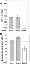
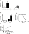
Adult schistosomes are intravascular parasites that metabolize imported glucose largely via glycolysis. How the parasites get rid of the large amounts of lactic acid this generates is unknown at the molecular level. Here, we report that worms whose aquaporin gene (SmAQP) has been suppressed using RNAi fail to rapidly acidify their culture medium and excrete less lactate compared to controls. Functional expression of SmAQP in Xenopus oocytes demonstrates that this protein can transport lactate following Michaelis-Menten kinetics with low apparent affinity (Km = 41+/-5. 8 mM) and with a low energy of activation (E(a) = 7.18+/-0.7 kcal/mol). Phloretin, a known inhibitor of lactate release from schistosomes, also inhibits lactate movement in SmAQP-expressing oocytes. In keeping with the substrate promiscuity of other aquaporins, SmAQP is shown here to be also capable of transporting water, mannitol, fructose and alanine but not glucose. Using immunofluorescent and immuno-EM, we confirm that SmAQP is localized in the tegument of adult worms. These findings extend the proposed functions of the schistosome tegument beyond its known capacity as an organ of nutrient uptake to include a role in metabolic waste excretion.
Conflict of interest statement
Figures


Similar articles
-
The role of tegumental aquaporin from the human parasitic worm, Schistosoma mansoni, in osmoregulation and drug uptake.FASEB J. 2009 Aug;23(8):2780-9. doi: 10.1096/fj.09-130757. Epub 2009 Apr 13. FASEB J. 2009. PMID: 19364765 Free PMC article.
-
Metabolite movement across the schistosome surface.J Helminthol. 2012 Jun;86(2):141-7. doi: 10.1017/S0022149X12000120. Epub 2012 Feb 27. J Helminthol. 2012. PMID: 22365312 Review.
-
Schistosoma mansoni: biochemical characterization of lactate transporters or similar proteins.Exp Parasitol. 2006 Nov;114(3):180-8. doi: 10.1016/j.exppara.2006.03.007. Epub 2006 May 8. Exp Parasitol. 2006. PMID: 16682030
-
Amino acid transport in schistosomes: Characterization of the permeaseheavy chain SPRM1hc.J Biol Chem. 2007 Jul 27;282(30):21767-75. doi: 10.1074/jbc.M703512200. Epub 2007 Jun 1. J Biol Chem. 2007. PMID: 17545149
-
Fifty years of the schistosome tegument: discoveries, controversies, and outstanding questions.Int J Parasitol. 2021 Dec;51(13-14):1213-1232. doi: 10.1016/j.ijpara.2021.11.002. Epub 2021 Nov 9. Int J Parasitol. 2021. PMID: 34767805 Review.
Cited by
-
Identification and characterization of paramyosin from cyst wall of metacercariae implicated protective efficacy against Clonorchis sinensis infection.PLoS One. 2012;7(3):e33703. doi: 10.1371/journal.pone.0033703. Epub 2012 Mar 21. PLoS One. 2012. PMID: 22470461 Free PMC article.
-
A novel, non-neuronal acetylcholinesterase of schistosome parasites is essential for definitive host infection.Front Immunol. 2023 Jan 31;14:1056469. doi: 10.3389/fimmu.2023.1056469. eCollection 2023. Front Immunol. 2023. PMID: 36798133 Free PMC article.
-
Exploring the function of protein kinases in schistosomes: perspectives from the laboratory and from comparative genomics.Front Genet. 2014 Jul 31;5:229. doi: 10.3389/fgene.2014.00229. eCollection 2014. Front Genet. 2014. PMID: 25132840 Free PMC article.
-
Structural and evolutionary divergence of aquaporins in parasites (Review).Mol Med Rep. 2017 Jun;15(6):3943-3948. doi: 10.3892/mmr.2017.6505. Epub 2017 Apr 25. Mol Med Rep. 2017. PMID: 28440467 Free PMC article. Review.
-
The Role of Natural Products in Drug Discovery and Development against Neglected Tropical Diseases.Molecules. 2016 Dec 31;22(1):58. doi: 10.3390/molecules22010058. Molecules. 2016. PMID: 28042865 Free PMC article. Review.
References
-
- Gryseels B, Polman K, Clerinx J, Kestens L. Human schistosomiasis. Lancet. 2006;368:1106–1118. - PubMed
-
- Skelly PJ, Shoemaker CB. Induction cues for tegument formation during the transformation of Schistosoma mansoni cercariae. Int J Parasitol. 2000;30:625–631. - PubMed
-
- Skelly PJ, Shoemaker CB. The Schistosoma mansoni host-interactive tegument forms from vesicle eruptions of a cyton network. Parasitology. 2001;122 Pt 1:67–73. - PubMed
-
- Horemans AM, Tielens AG, van den Bergh SG. The transition from an aerobic to an anaerobic energy metabolism in transforming Schistosoma mansoni cercariae occurs exclusively in the head. Parasitology. 1991;102 Pt 2:259–265. - PubMed
-
- Tielens AG, Verwijs A, Elfring RH, van den Heuvel JM, van den Bergh SG. Schistosoma mansoni: rapid turnover of glycogen by adult worms in vivo. Exp Parasitol. 1989;68:235–237. - PubMed
Publication types
MeSH terms
Substances
Grants and funding
LinkOut - more resources
Full Text Sources

