Successful endoscopic injection sclerotherapy of high-risk gastroesophageal varices in a cirrhotic patient with hemophilia A
- PMID: 20454701
- PMCID: PMC2862315
- DOI: 10.1155/2010/518260
Successful endoscopic injection sclerotherapy of high-risk gastroesophageal varices in a cirrhotic patient with hemophilia A
Abstract
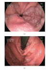
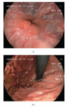
A 68-year-old man with hemophilia A and liver cirrhosis caused by hepatitis C virus was referred to our hospital to receive prophylactic endoscopic treatment for gastroesophageal varices (GOV). He had large, tense, and winding esophageal varices (EV) with cherry red spots extending down to lesser curve, predicting the likelihood of bleeding. Esophageal endoscopic injection sclerotherapy (EIS) was performed with a total 15 mL of 5% ethanolamine oleate with iopamidol (EOI). Radiographic imaging during EIS demonstrated that 5% EOI reached the afferent vein of the varices. He was administered sufficient factor VIII concentrate before and after EIS to prevent massive bleeding from the varices. Seven days after EIS, upper gastrointestinal endoscopy (UGIE) showed that the varices were eradicated almost completely. Eighteen months after EIS, the varices continued to diminish. We report a successful case of safe and effective EIS for GOV in a high-risk cirrhotic patient with hemophilia A.
Figures


Similar articles
-
Balloon-compression endoscopic injection sclerotherapy for the treatment of esophageal varices.VideoGIE. 2021 Oct 22;7(1):23-25. doi: 10.1016/j.vgie.2021.09.008. eCollection 2022 Jan. VideoGIE. 2021. PMID: 35059535 Free PMC article.
-
Hemodynamic evaluation by endoscopic ultrasonography of esophageal varices resistant to injection sclerotherapy.J Med Ultrason (2001). 2008 Mar;35(1):19-25. doi: 10.1007/s10396-007-0165-8. Epub 2008 Mar 15. J Med Ultrason (2001). 2008. PMID: 27278560
-
Evaluation of endoscopic injection sclerotherapy with and without simultaneous ligation for the treatment of esophageal varices.J Gastroenterol. 1999 Apr;34(2):159-62. doi: 10.1007/s005350050237. J Gastroenterol. 1999. PMID: 10213112 Clinical Trial.
-
Endoscopic variceal ligation compared with endoscopic injection sclerotherapy for treatment of esophageal variceal hemorrhage: a meta-analysis.World J Gastroenterol. 2015 Feb 28;21(8):2534-41. doi: 10.3748/wjg.v21.i8.2534. World J Gastroenterol. 2015. PMID: 25741164 Free PMC article. Review.
-
Endoscopic sclerotherapy.Surg Clin North Am. 1990 Apr;70(2):341-59. doi: 10.1016/s0039-6109(16)45085-1. Surg Clin North Am. 1990. PMID: 2181708 Review.
References
-
- Goedert JJ, Eyster ME, Lederman MM, et al. End-stage liver disease in persons with hemophilia and transfusion-associated infections. Blood. 2002;100(5):1584–1589. - PubMed
-
- Sarin SK, Lahoti D, Saxena SP, Murthy NS, Makwana UK. Prevalence, classification and natural history of gastric varices: a long-term follow-up study in 568 portal hypertension patients. Hepatology. 1992;16(6):1343–1349. - PubMed
-
- Ryan BM, Stockbrugger RW, Ryan JM. A pathophysiologic, gastroenterologic, and radiologic approach to the management of gastric varices. Gastroenterology. 2004;126(4):1175–1189. - PubMed
-
- World Federation of Hemophilia. Guidelines for the Management of Hemophilia. 2005.
-
- Furie B, Limentani SA, Rosenfield CG. A practical guide to the evaluation and treatment of hemophilia. Blood. 1994;84(1):3–9. - PubMed
Publication types
LinkOut - more resources
Full Text Sources

