. 2010 Jun 14;12(22):5861-7.
doi: 10.1039/c003763j. Epub 2010 May 8.

Resolution and polarization distribution in cryogenic DNP/MAS experiments

Affiliations

Resolution and polarization distribution in cryogenic DNP/MAS experiments

Alexander B Barnes et al. Phys Chem Chem Phys. .

Abstract

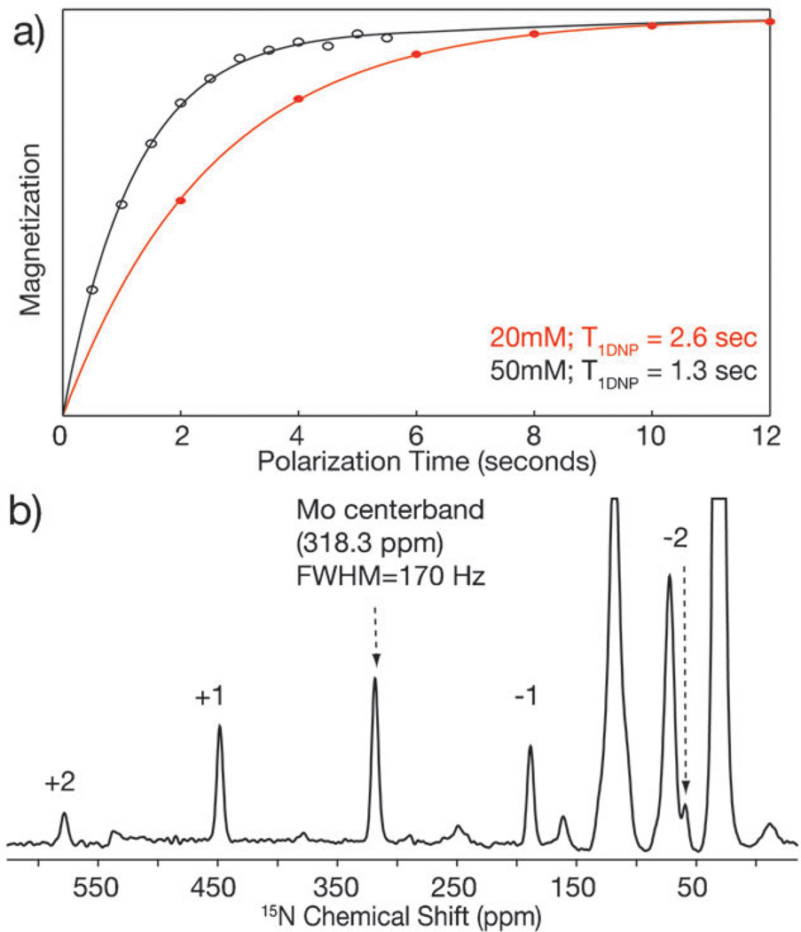
This contribution addresses four potential misconceptions associated with high-resolution dynamic nuclear polarization/magic angle spinning (DNP/MAS) experiments. First, spectral resolution is not generally compromised at the cryogenic temperatures at which DNP experiments are performed. As we demonstrate at a modest field of 9 T (380 MHz (1)H), 1 ppm linewidths are observed in DNP/MAS spectra of a membrane protein in its native lipid bilayer, and <0.4 ppm linewidths are reported in a crystalline peptide at 85 K. Second, we address the concerns about paramagnetic broadening in DNP/MAS spectra of proteins by demonstrating that the exogenous radical polarizing agents utilized for DNP are distributed in the sample in such a manner as to avoid paramagnetic broadening and thus maintain full spectral resolution. Third, the enhanced polarization is not localized around the polarizing agent, but rather is effectively and uniformly dispersed throughout the sample, even in the case of membrane proteins. Fourth, the distribution of polarization from the electron spins mediated via spin diffusion between (1)H-(1)H strongly dipolar coupled spins is so rapid that shorter magnetization recovery periods between signal averaging transients can be utilized in DNP/MAS experiments than in typical experiments performed at ambient temperature.


Figures

Fig. 1
Fig. 1
High resolution DNP spectra of bR. (a) RFDR correlation spectrum of 14 mg of [14–13C]retinal, [ε-13C]lysine-labeled bR in the dark-adapted state with 1D direct 13C dimension slices overlaid on the 2D contours. 12.5 days worth of acquisition time from mixing times between 8 and 28 ms were averaged together. The DNP enhancement is ~40, relative to the signal recorded in the absence of microwaves. ωr/2π = 8000 Hz and the temperature is 93 K. The resulting signal-to-noise negated the need for any line-broadening. The bR568 correlation in the upper left has a linewidth of 1.2 ppm (115 Hz). (b) Expansion of the lower right portion of the spectrum in (a) demonstrating the narrow linewidths achievable with MAS DNP. Whereas the bR555 correlation is slightly broader due to the presence of two conformations of bR555, the bR568 resonance is extremely narrow with a linewidth of 1.0 ppm (95 Hz). (c) Carbon–nitrogen 2D spectrum of uniformly 13C, 15N-labeled bR568, ωr/2π = 6000 Hz. After an indirect chemical shift evolution on the resolved Schiff base (165 ppm in the 15N dimension), the magnetization is transferred along the retinal chain all the way to C11, and also in the other direction directly to Cε. The carbon resonances show relatively narrow linewidths (1.2–2.2 ppm).
Fig. 2
Fig. 2
Temperature dependent high-resolution MAS spectra of N-f-MLF-OH. The spinning frequency is between 4000 and 5500 Hz, with −5 Hz stability. (a) Temperature dependent 13C spectra from 273 to 75 K showing that excellent resolution is maintained throughout the 200 K temperature range. (b) Expansion of the aromatic regions at 273, 190, and 82 K showing resolved resonances, even with coexisting conformations at 190 K. (c) Expansion of the alpha carbon region showing the resolved resonances of the two conformations present at 75 K, each different from the resonances of the single conformation observed at 273 K. (d) 15N spectrum of [U-13C, 15N]-N-f-MLF-OH. The shoulder on the resonances is a manifestation of the two conformations present at 75 K. Nonetheless, excellent resolution (linewidth of 39 Hz) is still maintained at 75 K.
Fig. 3
Fig. 3
High-resolution spectra of APG showing low temperature heterogeneous broadening for only the 3 carbon resonances that sample two puckered conformations, ωr/2π = 4831 Hz. (a) 13C spectra of APG at 293 and 75 K. (b) The carbon aliphatic region of a 15N–13C correlation spectrum with 1D direct 13C dimension slices overlaid on the 2D contours. (c) X-Ray and solid state NMR structures of APG from Barnes et al.
Fig. 4
Fig. 4
1D 15N spectrum of U-[13C, 15N]-bR showing the 15N Schiff base resonance adjacent to the peptide backbone signal, ωr/2π = 6000 Hz.
Fig. 5
Fig. 5
Accelerated DNP (a) difference in longitudinal relaxation time, T1DNP, under microwave irradiation between 20 and 50 mM TOTAPOL. The magnetization of the bulk protons is measured via CP to the carbonyls of [U-15N, 13C]-bR. (b) DNP spectrum of [ζ-15N]lysine-labeled bR trapped in the Mo photocycle intermediate. 81 920 Transients, 1.6 s recycle delay, 36.4 h acquisition time, ωr/2π = 5000 Hz, T = 89 K. The isotropic shift is 318.3 ppm, the span is 620 ppm, the skew is −0.034, and the tensor elements are: δ11 = 618, δ22 = 340, and δ33 = −2.5 ppm.

Similar articles

Cited by

References

    1. Carver TR, Slichter CP. Phys. Rev. 1956;102:975–980.
    1. Rosay M, Lansing JC, Haddad KC, Bachovchin WW, Herzfeld J, Temkin RJ, Griffin RG. J. Am. Chem. Soc. 2003;125:13626–13627. - PubMed
    1. Barnes AB, De Paëpe G, van der Wel P, Hu K, Joo C, Bajaj VS, Mak-Jurkauskas M, Sirigiri JR, Herzfeld J, Temkin R, Griffin RG. Appl. Magn. Reson. 2008;34:237–263. - PMC - PubMed
    1. Mak-Jurkauskas ML, Bajaj VS, Hornstein MK, Belenky M, Temkin RJ, Griffin RG, Herzfeld J. Proc. Natl. Acad. Sci. U. S. A. 2008;105:883–888. - PMC - PubMed
    1. Debelouchina GT, Bayro MJ, van der Wel PCA, Caproini M, Barnes AB, Rosay M, Werner M, Griffin RG. Phys. Chem. Chem. Phys. 2010 - PMC - PubMed

Publication types