

Review
doi: 10.1007/s00018-010-0390-y.
Epub 2010 May 9.
Glial versus melanocyte cell fate choice: Schwann cell precursors as a cellular origin of melanocytes
Affiliations
- PMID: 20454996
- PMCID: PMC11115498
- DOI: 10.1007/s00018-010-0390-y
Item in Clipboard
Review
Glial versus melanocyte cell fate choice: Schwann cell precursors as a cellular origin of melanocytes
Cell Mol Life Sci.
2010 Sep.
Abstract
Melanocytes and Schwann cells are derived from the multipotent population of neural crest cells. Although both cell types were thought to be generated through completely distinct pathways and molecular processes, a recent study has revealed that these different cell types are intimately interconnected far beyond previously postulated limits in that they share a common post-neural crest progenitor, i.e. the Schwann cell precursor. This finding raises interesting questions about the lineage relationships of hitherto unrelated cell types such as melanocytes and Schwann cells, and may provide clinical insights into mechanisms of pigmentation disorders and for cancer involving Schwann cells and melanocytes.
Figures

Two origins of melanocytes in the embryo: NCCs and SCPs from peripheral innervation. a Section through the E4 chick embryo at the forelimb level. MEBL1 and MITF (markers for melanocytes) are labeled in green and red respectively, Sox10 (SCP and melanoblasts marker) is shown in a blue color. Note the NCC-derived melanoblasts labeled with MEBL-1 and MITF at the dorsal skin above the neural tube. Melanoblasts emerging from SCPs are adjacent to the ventral branch of spinal nerve (vbSN) and are spatially separated from the population of NCC-derived melanocytes. DRG, dorsal root ganglion; NT, neural tube. b 3D representation outlining two separate pathways of melanocytes at the forelimb level in avian embryo. Red color represents melanoblasts, green color, nerves; purple, dermamyotome. c Scheme illustrating the relationship between SCPs, melanocytes and neurons as a result of NC differentiation. Conversion of SCPs into melanocytes is shown by the red arrow. In this case SCPs could correspond to the classical glia-melanocyte precursor postulated in earlier work by Elisabeth Dupin and Nicole M. Le Douarin [1, 97]. d Section with melanoblast adjacent to the dorsal ramus of spinal nerve. Nerve fibers are labeled in green, Sox10+ nuclei of SCPs and melanoblasts are shown in blue, and the red stands for melanocyte marker DCT. e Scheme showing the same cell biased by competition between signaling from the nerve (in a blue frame) and other paracrine signals promoting a melanocyte fate (in a red frame). f Identification of SCP-derived melanocytes in hair follicles and in dermis of adult mice by a genetic tracing approach. PLP-CreERT2 mouse strain was crossed with Rosa26stopYFP strain, and pregnant females were injected with tamoxifen (TM) at the post-NC stage (E11). Subsequent analysis identified numerous YFP+ cells among the population of dermal solitude melanocytes (DM) and in melanocytes of the hair follicles (HFM) in PLP-CreERT2—Rosa26stopYFP mice
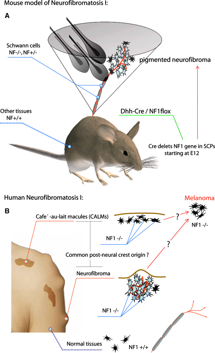
Common post-NC origin of melanocytes and tumorigenic SCs in neurofibromatosis type 1 disease. a Mouse model of neurofibromatosis based on the Dhh-Cre/NF1floxed mouse strain. Dhh promoter activates Cre expression in SCPs starting at E12–E12.5 causing deletion of the floxed NF1 gene. Later these mice develop pigmented neurofibroma tumors. b Neurofibromatosis type 1 in humans. Melanocytes in café-au-lait macules (CALMs) are deficient in NF1 gene on the background of normal melanocytes in the rest of the body. Patients with both neurofibromatosis type 1 and melanoma show biallelic deletion of NF1 in tumorigenic melanocytes. These data inspired us to propose a hypothesis implying common post-NC origin of melanocytes in pigmentation abnormalities together with melanocytes in melanoma tumors and mosaic tumorigenic SCs of neurofibromatosis type 1 patients
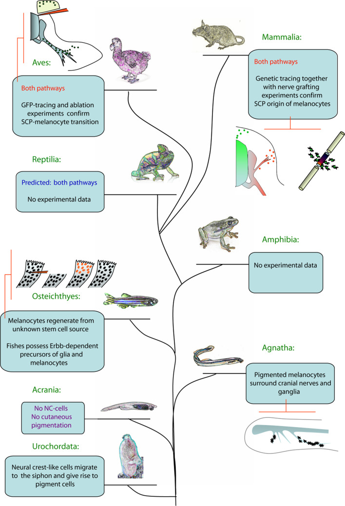
Emergence of the nerve-derived pathway of melanocytes in evolution. This evolutionary tree holds landmarks showing different phenomena putatively related to the conversion of SCPs into melanocytes. Pigmentation of cranial nerves in cyclostome larva and the presence of enigmatic ErbB-dependent nonpigmented melanocyte precursors in zebrafish are good examples of preliminary data showing that nerve-derived melanocytes could potentially appear at one of those levels of organization. Pictograms outline the crucial experiments and observations corroborating the aforementioned idea. Mammalia: on the left pictogram, green symbolizes the NCC-derived melanoblasts with origin in the neural tube (gradient green), and red outlines are melanoblasts derived from SCPs positioned along the nerve (gradient red). The right pictogram depicts the nerve transection experiment (grafted piece is labeled with dark color surrounded by newly formed melanocytes). The Aves pictogram depicts the experiment with surgical ablation of the NC-derived pathway of melanoblasts combined with GFP tracing of NC derivatives. GFP melanoblasts (small circles) are found later in the limb of the embryos with previously deleted dorsal skin and dermamyotome. Osteichtyes; red symbolizes newly formed melanocytes after fin ablation in zebrafish. Melanin does not redistribute in those cells in the presence of PTU, confirming their alternative origin. Agnatha: the nervous system (cranial nerves) of the cyclostome larva is marked by grey color, pigmented melanocytes are black
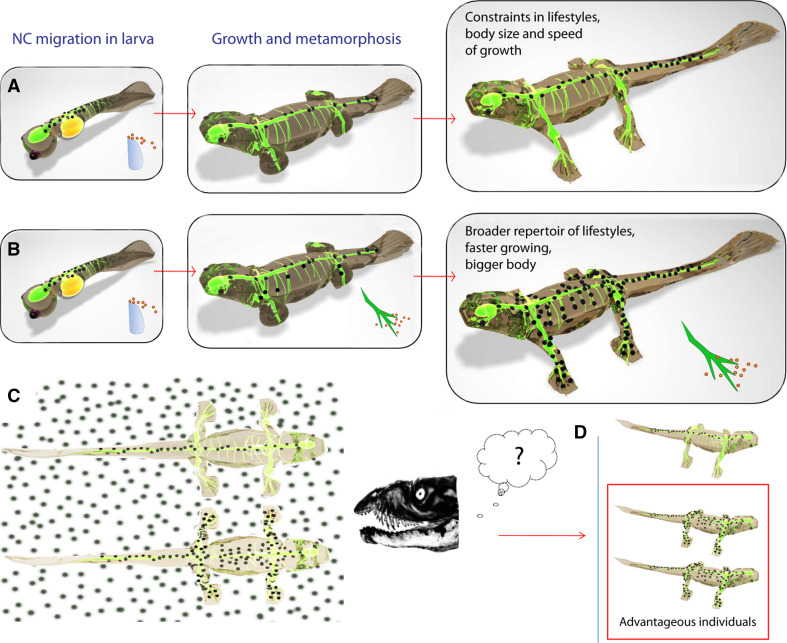
Advantage of a hypothetical vertebrate possessing the nerve-derived pathway of melanocytes compared with a fellow creature with only NC-derived melanocytes. a Development and pigmentation of imaginary vertebrate with NC-derived melanocytes only. b Development and pigmentation of imaginary vertebrate with both pathways generating primary NC-derived melanocytes and later nerve-derived melanocytes. c Insufficient pigmentation is disadvantageous to such individuals (shown in a) faced with active predators. d Presence of both mechanisms producing melanocyte progenitors ensures better camouflage and survival. a–d
Green color outlines the nervous system of an animal. Developmental strategies of living creatures always meet environmental criteria and constraints for better fitting and survival. Even tiny growing animals, larvae, should be safe during their growth and maturation. For this reason the speed of their development and their body size should be dependent on their ability to become fully pigmented at the proper time. Furthermore, the number of melanocyte progenitors generated at such early stages must be sufficient to provide a pigmentation pattern of an adult animal later. Insufficiently pigmented individuals are easily eliminated by predators, and, solely because of this, they must hide their bodies, acquire benthic way of life, their activity becomes restricted to the dark part of the day, they are forced to adapt to very deep or permanently dark habitats, for example caves. However, acquisition of mechanisms providing the animal with nerve-derived melanocytes could solve these tricky constraints, giving more developmental flexibility to the system. Animals possessing both ways of making melanocytes can gain faster growth rates and bigger bodies, solely because the speed of larval growth no longer depends on the migration of initial melanocyte precursors. Instead, new precursors can be recruited locally, providing the animal with specific pigmentation pattern. Hence, the lifestyle of the animal could potentially reflect the contribution of each pathway of melanocytes. We could also expect that the nerve-derived pathway of melanocytes appeared in evolution earlier than vertebrate embryos acquired protecting shells
References
-
- Adameyko I, Lallemend F, Aquino JB, Pereira JA, Topilko P, Muller T, Fritz N, Beljajeva A, Mochii M, Liste I, Usoskin D, Suter U, Birchmeier C, Ernfors P. Schwann cell precursors from nerve innervation are a cellular origin of melanocytes in skin. Cell. 2009;139:366–379. doi: 10.1016/j.cell.2009.07.049. - DOI - PubMed
Publication types
MeSH terms
LinkOut - more resources
Full Text Sources
Medical
