. 2011 Jan;101(1):1-14.
doi: 10.1007/s11060-010-0220-y. Epub 2010 May 9.

Knockdown of CypA inhibits interleukin-8 (IL-8) and IL-8-mediated proliferation and tumor growth of glioblastoma cells through down-regulated NF-κB

Affiliations

Knockdown of CypA inhibits interleukin-8 (IL-8) and IL-8-mediated proliferation and tumor growth of glioblastoma cells through down-regulated NF-κB

Shan Sun et al. J Neurooncol. 2011 Jan.

Abstract

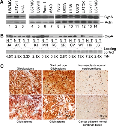
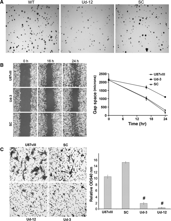
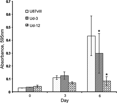
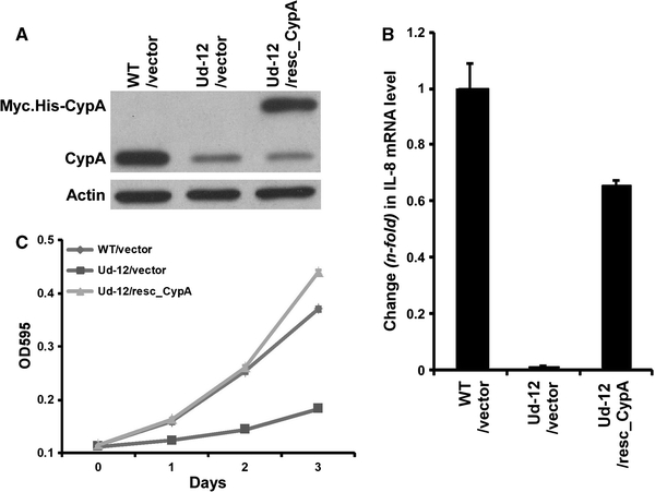
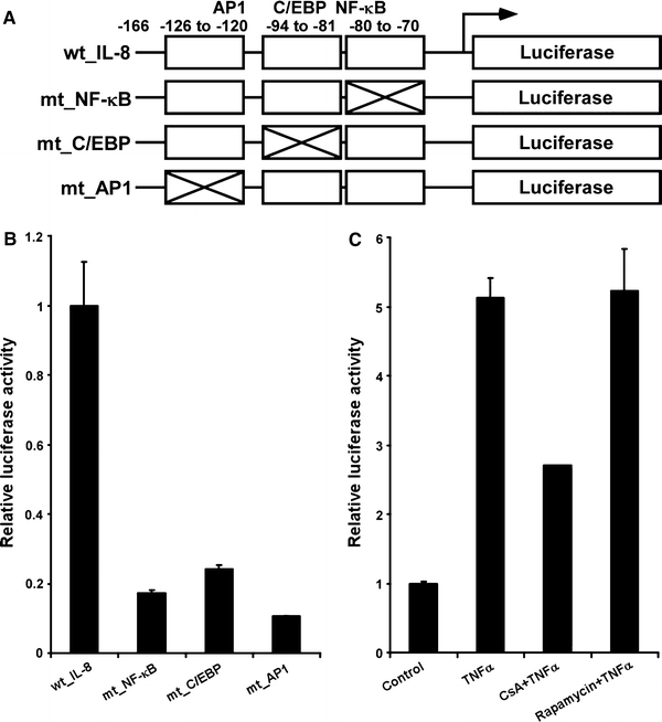
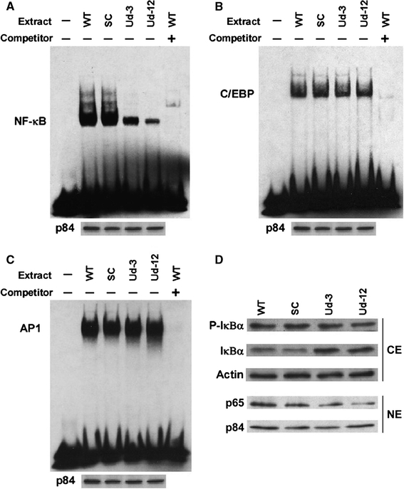
Although cyclophilin A (CypA) has been reported to be over-expressed in cancer cells and solid tumors, its expression and role in glioblastomas have not been studied. Herein, we show that expression of CypA in human glioblastoma cell lines and tissues is significantly higher than in normal human astrocytes and normal counterparts of brain tissue. To determine the role of over-expressed CypA in glioblastoma, stable RNA interference (RNAi)-mediated knockdown of CypA (CypA KD) was performed in gliobastoma cell line U87vIII (U87MG · ΔEGFR). CypA KD stable single clones decrease proliferation, infiltration, migration, and anchorage-independent growth in vitro and with slower growth in vivo as xenografts in immunodeficient nude mice. We have also observed that knockdown of CypA inhibits expression of interleukin-8 (IL-8), a tumorigenic and proangiogenic cytokine. Conversely, enforced expression of CypA in the CypA KD cell line, Ud-12, markedly enhanced IL-8 transcripts and restored Ud-12 proliferation, suggesting that CypA-mediated IL-8 production provides a growth advantage to glioblastoma cells. CypA knockdown-mediated inhibition of IL-8 is due to reduced activity of NF-κB, which is one of the major transcription factors regulating IL-8 expression. These results not only establish the relevance of CypA to glioblastoma growth in vitro and in vivo, but also suggest that small interfering RNA-based CypA knockdown could be an effective therapeutic approach against glioblastomas.


Figures

Fig. 1
Fig. 1
Over-expression of CypA in glioblastoma cell lines and human tissue specimens. a Elevated CypA at the protein level in glioblastoma cell lines. Five micrograms of cell lysate prepared from U87MG and U87MG. ΔEGFR (U87vIII) and normal human astrocyte (NHA) were subjected to Western blot analysis to detect CypA expression levels (lanes 1–3). Actin was used as a loading control. Over-expression of CypA levels in glioblastoma cell lines is comparable with those in pancreatic and non-small cell lung carcinoma cell lines (lanes 6 and 7). Lanes 8–11 are other glioblastoma cell lines, and U87DK (lane 12) represents K721 mutation and kinase inactive at EGFR. b Relative CypA levels in human glioblastoma and normal tissues. Ten micrograms of tissue lysates were subjected to Western blot analysis. Coomassie brilliant blue R250-stained signals on the nitrocellulose membrane served as internal controls. (N, normal brain tissue; T, tumor tissue). c CypA expression in glioblastoma multiforms (GBMs). Deparaffinized and hydrated tissue sections were hybridized with rabbit anti-cyclophilin A antibody (Abcam), followed by incubating sections with ImmPress anti-rabbit Ig (Vector Lab) and DAB (brown) staining. Arrows indicate typical positive staining for CypA. Non-neoplastic normal cerebrum tissue and cancer-adjacent normal cerebrum tissue were used as controls
Fig. 2
Fig. 2
CypA KD single clones result in decreased cell proliferation and tumor growth. a CypA KD stable single clones generated from U87vIII cells. U87vIII cells were seeded into 24-well plates at 5 × 104 cells per well 1 day before infection with enriched viral media prepared from the CypA-specific siRNA insert, scrambled siRNA vector, and linearized retrovirus vector (supplied with the pSilencer H1-Retro kit purchased from Ambion). Cells were transferred into 6-well plates 3 days post-infection and selected with 2 μg/ml puromycin for 2 weeks. Puromycin-resistant clones were harvested and lysed with RIPA buffer supplemented with a protease inhibitor cocktail (Sigma). Five micrograms of total protein was used for Western blot analysis. Membranes probed with actin antibody were used as loading controls. b CypA KD results in decreased cell proliferation. Ud-3 and Ud-12 were compared to parental U87vIII cells and scrambled clone (SC), using MTT assays according to the manufacturer’s protocol. Values of OD 595 absorbance at each time point were the average of triplicate individual readings (bars, SE; *, P < 0.05 compared with parental wild-type cells). c CypA KD decreases tumor growth. Taconic Balb/c/NIH(s) female nu/nu mice were used for xenografts. One million cells were injected. At each time point, tumor dimensions were measured with calipers, and volume was calculated as V= 0.5 × L × W 2. Three mice were used at each time point and each cell type (bars, SE; *, P < 0.05 compared with parental wild-type cells)
Fig. 3
Fig. 3
The CypA KD U87vIII single clone decreases anchorage-independent growth, and reduces cell migration and infiltration ability in vitro. a Anchorage-independent growth in vitro. Ud-12 and parental U87vIII cells and SC were plated in duplicate in six-well plates in 0.5% soft agarose on a 1.4% agarose bed agar medium. After 21 days of incubation, the cells were stained with crystal violet and imaged. Colonies larger than 0.2 mm2 were counted. Ud-12/U87vIII= 47.6%, P < 0.05. b Knock down of CypA in U87vIII reduces cell migration. Ud-3 cells were seeded into 6-well plates. When cells reached subconfluent monolayers, a scratch wound was inflicted, and the resulting gap was imaged at the time of the wound and after 16 and 24 h. Images are from a typical experiment. c Infiltration assay in vitro. Transwell plates from Corning Life Sciences were used for invasion assays. The effect of CypA KD on cell infiltration was evaluated using Matrigel as an extracellular matrix component. Chambers were incubated for 24 h under normal cell culture conditions. Invading cells on the lower side of the filter were stained with 0.1% crystal violet (left panel) and quantified after dissolving the cell-bound crystal violet in 10% acetic acid (v/v), followed by measurement of optical density at 540 nm. (Columns, mean of the relative OD 540 nm readings from three independent experiments; bars, SE (right panel); #, P < 0.05 compared with parental wild-type cells)
Fig. 4
Fig. 4
CypA KD correlates with increased apoptosis under the condition of serum withdrawal. The CypA KD single clones, Ud-3 and Ud-12, and parental U87vIII control cells were grown in 96-well plates in media without serum for 6 days. Cells (10,000/well) were plated in replicates of three. Viable cells were measured at the indicated time points by MTT assays. Columns, mean of the relative OD 595 nm readings from three independent experiments; bars, SE; *, P < 0.05 compared with parental wild-type cells
Fig. 5
Fig. 5
CypA KD-mediated down-regulation of IL-8 expression. a IL-8 expression levels were determined by a quantitative real-time PCR analysis. One microgram of total RNA was used for cDNA synthesis, using Superscript III (Invitrogen) with the oligo(dT)15 primer. Quantitative real-time PCR (QRT-PCR) was performed using the Power SYBR Green PCR Master Mix (Applied Biosystem) detected by the 7300 Real Time PCR System (Applied Biosystems). The relative expression of IL-8 mRNA normalized to the internal reference 18S rRNA was analyzed using the 2−ΔΔCT method (columns, mean of results from three independent experiments; bars, SE; *, P < 0.05 compared with parental wild-type cells). b IL-8 ELISA. Cells were seeded into 24-well plates for 24 h. Cell media was collected for measurement of IL-8 expression according the manufacturer’s protocol (columns, mean of results from three independent experiments; bars, SE; *, P < 0.05 compared with parental wild-type cells)
Fig. 6
Fig. 6
Expression of a siRNA-resistant wild-type CypA rescue expression plasmid restores IL-8 expression and cell proliferation defects. a Western blot analysis of endogenous CypA and exogenous myc-tagged CypA. U87vIII cells (WT) and CypA KD clone Ud-12 were transfected with the pcDNA3.1/Myc.His vector (vector) or the rescue construct (resc_CypA). Five micrograms of each cell extract were subjected to Western blot analysis probing with anti-CypA and anti-myc antibodies. b Real-time PCR analysis of the IL-8 mRNA. The values plotted are the mean of three separate experiments. c MTT assay of cell proliferation. At various time points, cells were subjected to MTT assays. The absorbance in each well was measured at 595 nm in a microtiter plate reader. Bars indicate the standard error from three individual experiments
Fig. 7
Fig. 7
Analysis of IL-8 promoter and regulation of IL-8 expression by TNFα and CsA. a The schematic IL-8 luciferase reporter constructs used in this study. The luciferase reporter plasmids driven by wild-type IL-8 promoter and the mutated promoters containing mutated NF-κB, C/EBP, or AP1 site are designated as wt_IL-8, mt_NF-κB, mt_C/EBP, and mt_AP1, respectively. b U87vIII cells were transfected with indicated reporter plasmids together with Renilla luciferase plasmid. Luciferase activity was measured at 24 h post-transfection (columns, mean of results from three independent experiments; bars, SE). c U87vIII cells were transfected with the reporter plasmid driven by the wild type IL-8 promoter together with Renilla luciferase plasmid. At 24 h post-transfection, cells were incubated with 1 μg/ml CsA or rapamycin for 5 h, then stimulated by 10 ng/ml TNF-α for another 4 h. Cells were then harvested and promoter activity was determined. Results are presented as relative reporter activity after normalization to Renilla luciferase activity. Bars indicate the standard error of three replicates
Fig. 8
Fig. 8
Reduced NF-κB binding activity and decreased p65 associated with CypA KD cells. a, b, and c EMSA assays were performed using nuclear extracts from U87vIII cells (WT), scrambled control cells (SC), and the CypA KD clones, Ud-3 and Ud-12, with NF-κB, C/EBP, or AP1 oligos as probes. For competition experiments, protein was pre-incubated with 100-fold molar excess of unlabeled oligos at 25°C for 10 min before addition of labeled probes. p84 expression on Western blots was used as loading control for EMSA. d Cytoplasmic extracts (CE) prepared from U87vIII cells (WT), scrambled control cells (SC), and the CypA KD clones, Ud-3 and Ud-12, were immunoblotted for phosphorylated IκBα (p-IκBα) and IκBα, and actin was used as the internal control (upper panel). Nuclear extracts (NE) were subjected to Western blot analysis probing with anti-p65 antibody. p84 was used as an internal control (lower panel)

Similar articles

Cited by

References

    1. Gurney JG, Kadan-Lottick N. Brain and other central nervous system tumors: rates, trends, and epidemiology. Curr Opin Oncol. 2001;13:160–166. doi: 10.1097/00001622-200105000-00005. - DOI - PubMed
    1. Ross HJ, Cho J, Osann K, et al. Phase I/II trial of low dose cyclosporine A with EP for advanced non-small cell lung cancer. Lung Cancer. 1997;18:189–198. doi: 10.1016/S0169-5002(97)00061-5. - DOI - PubMed
    1. Sliwa M, Markovic D, Gabrusiewicz K, et al. The invasion promoting effect of microglia on glioblastoma cells is inhibited by cyclosporine A. Brain. 2007;130:476–489. doi: 10.1093/brain/awl263. - DOI - PubMed
    1. Zupanska A, Dziembowska M, Ellert-Miklaszewska A, et al. Cyclosporin A induces growth arrest or programmed cell death of human glioma cells. Neurochem Int. 2005;47:430–441. doi: 10.1016/j.neuint.2005.05.010. - DOI - PubMed
    1. Ryffel B, Woerly G, Greiner B, Haendler B, Mihatsch MJ, Foxwell BM. Distribution of the cyclosporine binding protein cyclophilin in human tissues. Immunology. 1991;72:399–404. - PMC - PubMed

MeSH terms