Comparative Study
. 2010 Sep;64(3):197-211.
doi: 10.1111/j.1600-0897.2010.00850.x.

Keratinocyte Growth Factor Stimulates Macrophage Inflammatory Protein 3α and Keratinocyte-derived Chemokine Secretion by Mouse Uterine Epithelial Cells

Affiliations
Comparative Study

Keratinocyte Growth Factor Stimulates Macrophage Inflammatory Protein 3α and Keratinocyte-derived Chemokine Secretion by Mouse Uterine Epithelial Cells

Severina N Haddad et al. Am J Reprod Immunol. 2010 Sep.

Abstract

Problem: communication between uterine epithelial cells and the underlying stromal fibroblasts is critical for proper endometrial function. Stromal fibroblast-derived growth factors have been shown to regulate epithelial immune functions. The purpose of this study was to determine whether keratinocyte growth factor (KGF) regulates uterine epithelial cell chemokine and antimicrobial secretion.

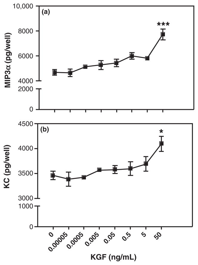
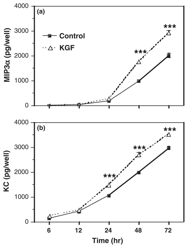
Method of study: uterine epithelial cells were isolated from Balb/c mice and cultured in either 96-well plates or transwell inserts. Epithelial cells were treated with KGF, epidermal growth factor (EGF), or hepatocyte growth factor (HGF). Macrophage inflammatory protein 3α (MIP3α) and keratinocyte-derived chemokine (KC) levels were measured by ELISA.

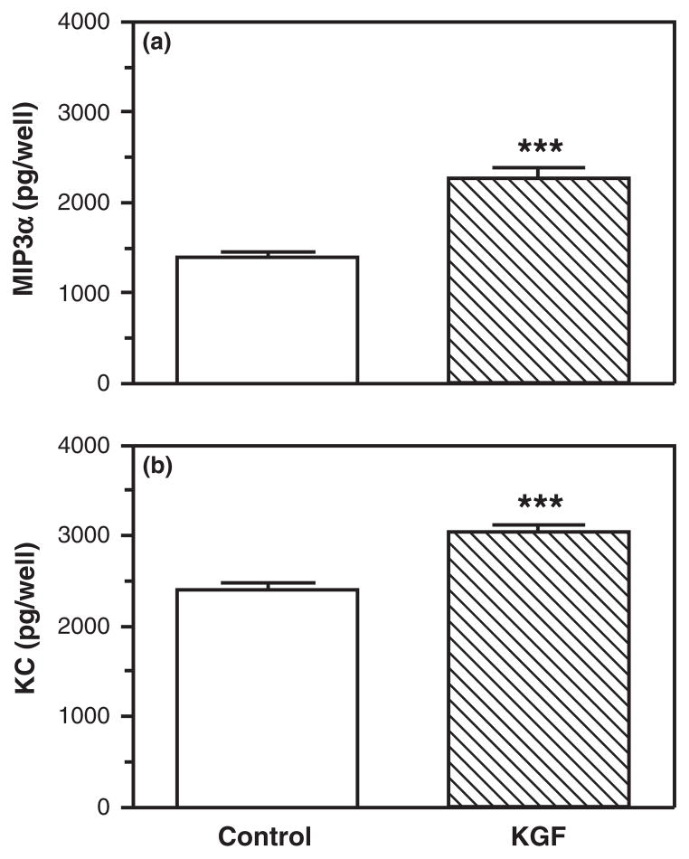
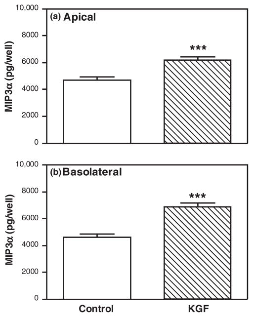
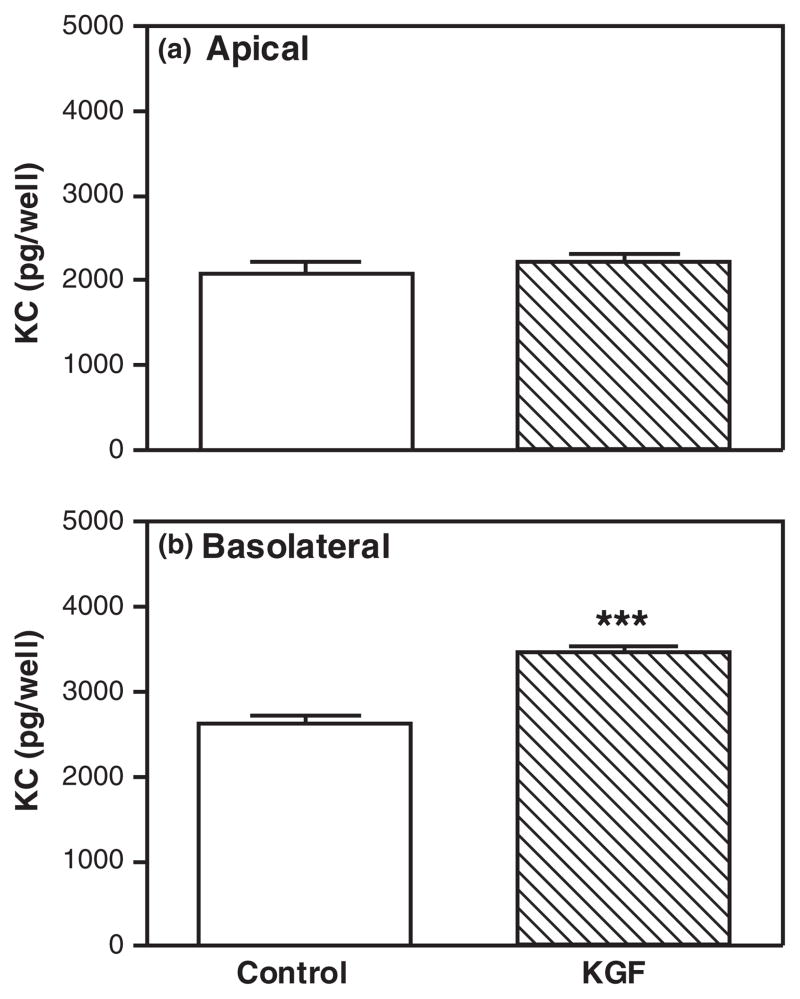
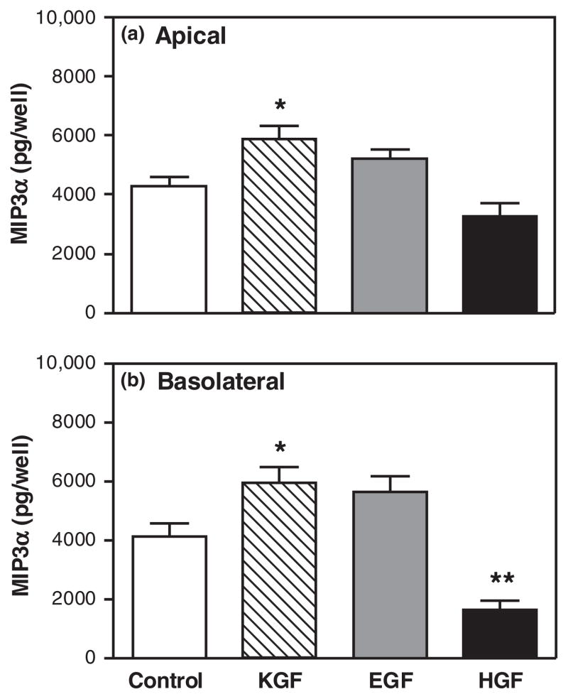
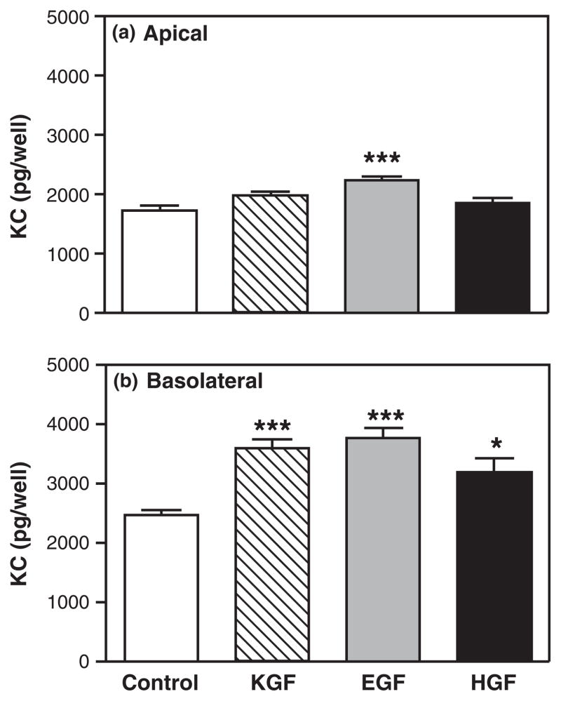
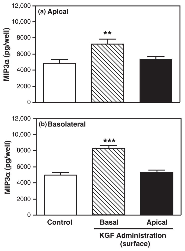
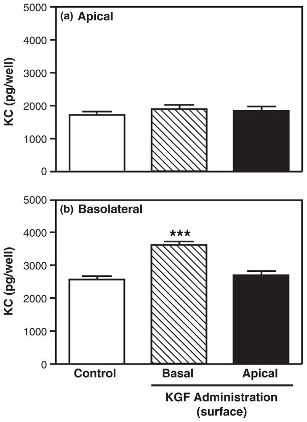
Results: keratinocyte growth factor stimulated the secretion of MIP3α and KC. The effects on MIP3α by KGF were specific because EGF and HGF had no effect. In contrast, KGF, EGF, and HGF had similar effects on KC. Furthermore, KGF administered to the apical side of epithelial cells had no effect on MIP3α or KC secretion, indicating that the KGF receptor is located on the basolateral surface of uterine epithelial cells.

Conclusion: we demonstrate that KGF plays a role in uterine epithelial cell secretion of MIP3α and KC, key immune mediators involved in the protection of mucosal surfaces in the female reproductive tract.


Figures

Fig. 1
Fig. 1
Keratinocyte growth factor (KGF) increases MIP3α and keratinocyte-derived chemokine (KC) secretion by freshly prepared mouse uterine epithelial cells. Freshly isolated mouse uterine epithelial cells were incubated overnight prior to treatment with control medium or KGF (50 ng/mL) for 48 hr. Supernatants were collected and analyzed for MIP3α (a) and KC (b). The results are shown as the mean + S.E.M. of eight separate experiments. ***MIP3α or KC significantly (P < 0.001) greater with KGF treatment than controls.
Fig. 2
Fig. 2
Keratinocyte growth factor (KGF) dose-dependently stimulates freshly prepared uterine epithelial cell release of MIP3α and keratinocyte-derived chemokine (KC). Freshly isolated mouse uterine epithelial cells were incubated overnight prior to treatment with KGF (0.00005–50 ng/mL) for 48 hr. Supernatants were collected and analyzed for MIP3α (a) and KC (b). The results are shown as the mean + S.E.M. ***MIP3α significantly (P < 0.001) or *KC significantly (P < 0.05) greater with KGF treatment than controls.
Fig. 3
Fig. 3
Time course of the effect of keratinocyte growth factor (KGF) on MIP3α and keratinocyte-derived chemokine (KC) secretion by uterine epithelial cells. Freshly isolated mouse uterine epithelial cells were incubated overnight prior to treatment with control medium or KGF (50 ng/mL) for 6, 12, 24, 48, or 72 hr. Supernatants were collected and analyzed for MIP3α (a) or KC (b). The results are shown as the mean + S.E.M. ***MIP3α or KC significantly (P < 0.001) greater with KGF treatment than controls.
Fig. 4
Fig. 4
Keratinocyte growth factor (KGF) increases apical and basolateral MIP3α secretion by polarized uterine epithelial cells. Mouse uterine epithelial cells were isolated and polarized on transwell cell culture inserts. Polarized epithelial cells were treated with control medium or KGF (50 ng/mL) in the basolateral compartment for 48 hr. Apical (a) and basolateral (b) supernatants were collected and analyzed for MIP3α. The results are shown as the mean + S.E.M. of 10 separate experiments. ***MIP3α significantly (P < 0.001) greater with KGF treatment than controls.
Fig. 5
Fig. 5
Keratinocyte growth factor (KGF) stimulates basolateral keratinocyte-derived chemokine (KC) secretion by polarized uterine epithelial cells. Mouse uterine epithelial cells were isolated and polarized on transwell cell culture inserts. Polarized epithelial cells were treated with control medium or KGF (50 ng/mL) in the basolateral compartment for 48 hr. Apical (a) and basolateral (b) supernatants were collected and analyzed for KC. The results are shown as the mean + S.E.M. of 10 separate experiments. ***KC significantly (P < 0.001) greater with KGF treatment than controls.
Fig. 6
Fig. 6
Specificity of keratinocyte growth factor (KGF) effect on MIP3α secretion. Mouse uterine epithelial cells were isolated and polarized on transwell cell culture inserts. Polarized epithelial cells were treated with control medium, KGF (50 ng/mL), epidermal growth factor (4 ng/mL), or HGF (50 ng/mL) in the basolateral compartment for 48 hr. Apical (a) and basolateral (b) supernatants were collected and analyzed for MIP3α. The results are shown as the mean + S.E.M. of four separate experiments. *MIP3α significantly (P < 0.05) greater with KGF treatment than with control medium. **MIP3α significantly (P < 0.01) less with HGF treatment than with control medium.
Fig. 7
Fig. 7
Specificity of keratinocyte growth factor (KGF) effect on keratinocyte-derived chemokine (KC) release. Mouse uterine epithelial cells were isolated and polarized on transwell cell culture inserts. Polarized epithelial cells were treated with control medium, KGF (50 ng/mL), epidermal growth factor (4 ng/mL), or HGF (50 ng/mL) in the basolateral compartment for 48 hr. Apical (a) and basolateral (b) supernatants were collected and analyzed for KC. The results are shown as the mean + S.E.M. of four separate experiments. *KC significantly (P < 0.05) greater with growth factor treatment than with control medium. ***KC significantly (P < 0.001) greater with growth factor treatment than with control medium.
Fig. 8
Fig. 8
Effect of keratinocyte growth factor (KGF) apical treatment on polarized uterine epithelial cell MIP3α secretion. Mouse uterine epithelial cells were isolated and polarized on transwell cell culture inserts. Polarized epithelial cells were treated with control medium, KGF (50 ng/mL) in the apical or basolateral compartment for 48 hr. Apical (a) and basolateral (b) supernatants were collected and analyzed for MIP3α. The results are shown as the mean + S.E.M. of two separate experiments. **MIP3α significantly (P < 0.01) greater with KGF treatment than with control medium. ***MIP3α significantly (P < 0.001) greater with KGF treatment than with control medium.
Fig. 9
Fig. 9
Effect of keratinocyte growth factor (KGF) apical treatment on keratinocyte-derived chemokine (KC) release by polarized uterine epithelial cells. Mouse uterine epithelial cells were isolated and polarized on transwell cell culture inserts. Polarized epithelial cells were treated with control medium, KGF (50 ng/mL) in the apical or basolateral compartment for 48 hr. Apical (a) and basolateral (b) supernatants were collected and analyzed for KC. The results are shown as the mean + S.E.M. of two separate experiments. ***KC significantly (P < 0.001) greater with KGF treatment than with control medium.

Similar articles

Cited by

References

    1. Janeway CA, Travers P, Walport M, Schlomchik MJ. Immunobiology: The Immune System in Health and Disease. 6. New York: Garland Science; 2005.
    1. Vaerman J-P. The secretory immune system. Antibiot Chemother. 1987;39:41–50. - PubMed
    1. Wira CR, Grant-Tschudy KS, Crane-Godreau MA. Epithelial cells in the female reproductive tract: a central role as sentinels of immune protection. Am J Reprod Immunol. 2005;53:65–76. - PubMed
    1. Wira CR, Fahey JV, Sentman CL, Pioli PA, Shen L. Innate and adaptive immunity in female genital tract: cellular responses and interactions. Immunol Rev. 2005;206:306–335. - PubMed
    1. Dockery P. The fine structure of the mature human endometrium. In: Glasser SR, Aplin JD, Giudice LC, Tabibzadeh S, editors. The Endometrium. New York: Taylor & Francis; 2002. pp. 21–38.

Publication types

MeSH terms