

Increased expression of bFGF is associated with carotid atherosclerotic plaques instability engaging the NF-κB pathway
- PMID: 20455997
- PMCID: PMC3822568
- DOI: 10.1111/j.1582-4934.2010.01082.x
Increased expression of bFGF is associated with carotid atherosclerotic plaques instability engaging the NF-κB pathway
Abstract
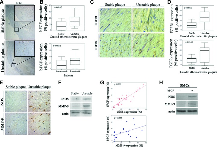
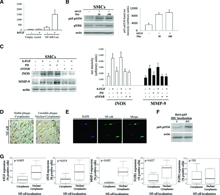
Unstable atherosclerotic plaques of the carotid arteries are at great risk for the development of ischemic cerebrovascular events. The degradation of the extracellular matrix by matrix metalloproteinases (MMPs) and NO-induced apoptosis of vascular smooth muscle cells (VSMCs) contribute to the vulnerability of the atherosclerotic plaques. Basic fibroblast growth factor (bFGF) through its mitogenic and angiogenic properties has already been implicated in the pathogenesis of atherosclerosis. However, its role in plaque stability remains elusive. To address this issue, a panel of human carotid atherosclerotic plaques was analyzed for bFGF, FGF-receptors-1 and -2 (FGFR-1/-2), inducible nitric oxide synthase (iNOS) and MMP-9 expression. Our data revealed increased expression of bFGF and FGFR-1 in VSMCs of unstable plaques, implying the existence of an autocrine loop, which significantly correlated with high iNOS and MMP-9 levels. These results were recapitulated in vitro by treatment of VSMCs with bFGF. bFGF administration led to up-regulation of both iNOS and MMP-9 that was specifically mediated by nuclear factor-kappaB (NF-kappaB) activation. Collectively, our data demonstrate a novel NF-kappaB-mediated pathway linking bFGF with iNOS and MMP-9 expression that is associated with carotid plaque vulnerability.
Figures
Similar articles
-
Basic fibroblast growth factor mediates carotid plaque instability through metalloproteinase-2 and -9 expression.Eur J Vasc Endovasc Surg. 2004 Jul;28(1):89-97. doi: 10.1016/j.ejvs.2004.02.022. Eur J Vasc Endovasc Surg. 2004. PMID: 15177237
-
Tumor necrosis factor-α regulates triggering receptor expressed on myeloid cells-1-dependent matrix metalloproteinases in the carotid plaques of symptomatic patients with carotid stenosis.Atherosclerosis. 2016 May;248:160-9. doi: 10.1016/j.atherosclerosis.2016.03.021. Epub 2016 Mar 19. Atherosclerosis. 2016. PMID: 27017522 Free PMC article.
-
Relation between TLR4/NF-κB signaling pathway activation by 27-hydroxycholesterol and 4-hydroxynonenal, and atherosclerotic plaque instability.Aging Cell. 2015 Aug;14(4):569-81. doi: 10.1111/acel.12322. Epub 2015 Mar 10. Aging Cell. 2015. PMID: 25757594 Free PMC article.
-
Potential role of insulin receptor isoforms and IGF receptors in plaque instability of human and experimental atherosclerosis.Cardiovasc Diabetol. 2018 Feb 20;17(1):31. doi: 10.1186/s12933-018-0675-2. Cardiovasc Diabetol. 2018. PMID: 29463262 Free PMC article.
-
The Role of Matrix Metalloproteinase-9 in Atherosclerotic Plaque Instability.Mediators Inflamm. 2020 Oct 6;2020:3872367. doi: 10.1155/2020/3872367. eCollection 2020. Mediators Inflamm. 2020. PMID: 33082709 Free PMC article. Review.
Cited by
-
C-reactive protein/oxidised low-density lipoprotein/β2-glycoprotein I complex promotes atherosclerosis in diabetic BALB/c mice via p38mitogen-activated protein kinase signal pathway.Lipids Health Dis. 2013 Mar 26;12:42. doi: 10.1186/1476-511X-12-42. Lipids Health Dis. 2013. PMID: 23531147 Free PMC article.
-
The association of functional polymorphisms in genes encoding growth factors for endothelial cells and smooth muscle cells with the severity of coronary artery disease.BMC Cardiovasc Disord. 2016 Nov 11;16(1):218. doi: 10.1186/s12872-016-0402-4. BMC Cardiovasc Disord. 2016. PMID: 27835972 Free PMC article.
-
bFGF blockade reduces intraplaque angiogenesis and macrophage infiltration in atherosclerotic vein graft lesions in ApoE3*Leiden mice.Sci Rep. 2020 Sep 29;10(1):15968. doi: 10.1038/s41598-020-72992-7. Sci Rep. 2020. PMID: 32994514 Free PMC article.
-
The underlying mechanisms of FGF2 in carotid atherosclerotic plaque development revealed by bioinformatics analysis.Arch Med Sci. 2021 May 9;20(4):1209-1219. doi: 10.5114/aoms/128387. eCollection 2024. Arch Med Sci. 2021. PMID: 39439688 Free PMC article.
-
Phenotypic Modulation of Macrophages and Vascular Smooth Muscle Cells in Atherosclerosis-Nitro-Redox Interconnections.Antioxidants (Basel). 2021 Mar 26;10(4):516. doi: 10.3390/antiox10040516. Antioxidants (Basel). 2021. PMID: 33810295 Free PMC article. Review.
References
-
- Sacco RL, Benjamin EJ, Broderick JP, et al. Risk factors: AHA Conference Proceedings. Stroke. 1997;28:1507–17. - PubMed
-
- Luigi Giusto S, Alessandro M, Giampiero P, et al. Relationships between risk factors and morphological patterns of human carotid atherosclerotic plaques. A multivariate discriminant analysis. Atherosclerosis. 1994;108:39–60. - PubMed
-
- Inzitari D, Eliasziw M, Gates P, et al. The causes and risk of stroke in patients with asymptomatic internal-carotid-artery stenosis. N Engl J Med. 2000;342:1693–701. - PubMed
-
- Sitzer M, Muller W, Siebler M, et al. Plaque ulceration and lumen thrombus are the main sources of cerebral microemboli in high-grade internal carotid artery stenosis. Stroke. 1995;26:1231–3. - PubMed
-
- Carr S, Farb A, Pearce WH, et al. Atherosclerotic plaque rupture in symptomatic carotid artery stenosis. J Vasc Surg. 1996;23:755–66. - PubMed
Publication types
MeSH terms
Substances
LinkOut - more resources
Full Text Sources
Medical
Miscellaneous
