

Cerebral white matter blood flow and arterial blood pressure in preterm infants
- PMID: 20456278
- PMCID: PMC3068289
- DOI: 10.1111/j.1651-2227.2010.01856.x
Cerebral white matter blood flow and arterial blood pressure in preterm infants
Abstract
It is generally assumed that one reason why white matter injury is common in preterm infants is the relatively poor vascular supply.
Aim: To examine whether blood flow to the white matter is relatively more reduced at low blood pressure than is blood flow to the brain as a whole.
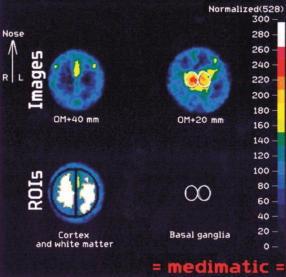
Methods: Thirteen normoxic preterm infants had blood flow imaging on 16 occasions with single-photon emission computed tomography (SPECT) using 99Tc labelled hexa-methylpropylenamide oxime (HMPAO) as the tracer. Gestational age was 26-32 weeks. Transcutaneous carbon dioxide was between 4.7 and 8.5 kPa and mean arterial blood pressure between 22 and 55 mmHg.
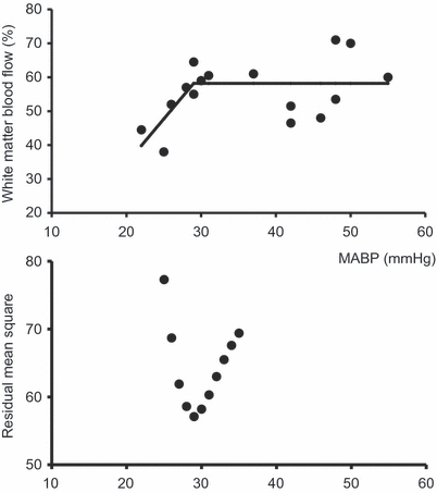
Results: There was no statistically significant direct relation between white matter blood flow percentage and any of the variables. Using non-linear regression, however, assuming a plateau over a certain blood pressure threshold and a positive slope below this threshold, the relation to white matter flow percentage was statistically significant (p = 0.02). The threshold was 29 mmHg (95% confidence limits 26-33).
Conclusion: Our analysis supports the concept of periventricular white matter as selectively vulnerable to ischaemia during episodes of low blood pressure.
© 2010 The Author(s)/Journal Compilation © 2010 Foundation Acta Paediatrica.
Figures
References
-
- Wigglesworth JS, Pape KE. An integrated model for haemorrhagic and ischaemic lesions in the newborn brain. Early Hum Dev. 1978;2:179–99. - PubMed
-
- Pryds O, Greisen G, Lou H, Friis-Hansen B. Heterogeneity of cerebral vasoreactivity in preterm infants supported by mechanical ventilation. J Pediatr. 1989;115:638–45. - PubMed
-
- Lou HC, Skov H, Pedersen H. Low cerebral blood flow: a risk factor in the neonate. J Pediatr. 1979;95:606–9. - PubMed
-
- Pryds O. Low neonatal cerebral oxygen delivery is associated with brain injury in preterm infants. Acta Paediatr. 1994;83:1233–6. - PubMed
MeSH terms
LinkOut - more resources
Full Text Sources
