Review
. 2010 Jul;217(1):1-15.
doi: 10.1111/j.1469-7580.2010.01227.x. Epub 2010 Apr 26.

Intervertebral disc, sensory nerves and neurotrophins: who is who in discogenic pain?

Affiliations
Review

Intervertebral disc, sensory nerves and neurotrophins: who is who in discogenic pain?

José García-Cosamalón et al. J Anat. 2010 Jul.

Abstract

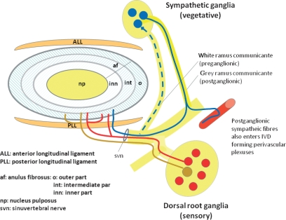
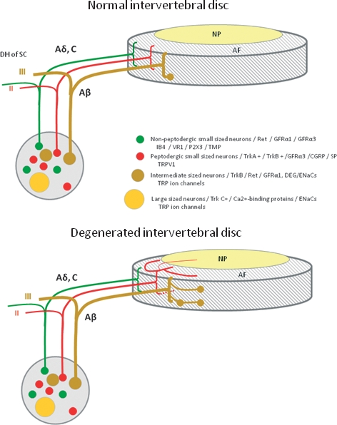
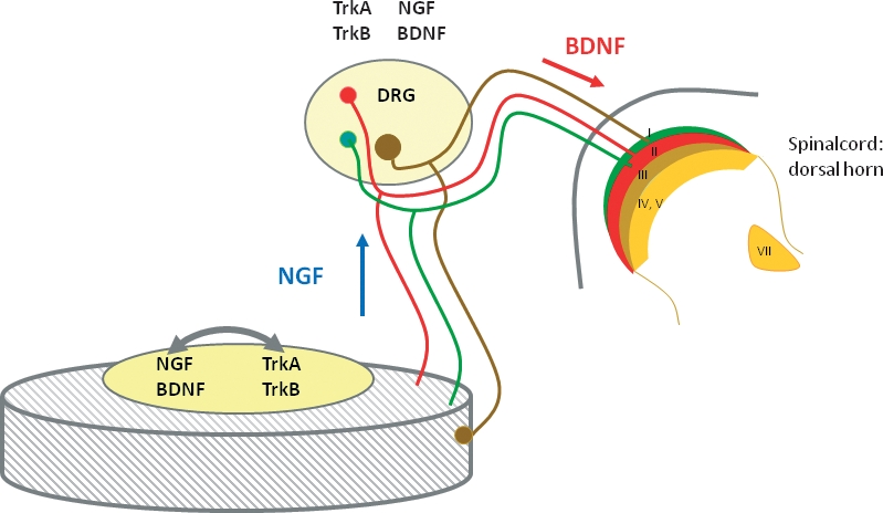
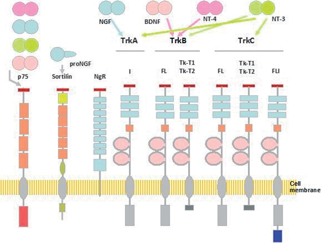
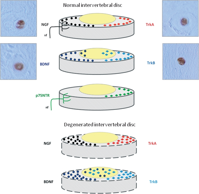
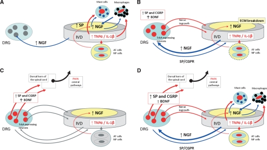
The normal intervertebral disc (IVD) is a poorly innervated organ supplied only by sensory (mainly nociceptive) and postganglionic sympathetic (vasomotor efferents) nerve fibers. Interestingly, upon degeneration, the IVD becomes densely innervated even in regions that in normal conditions lack innervation. This increased innervation has been associated with pain of IVD origin. The mechanisms responsible for nerve growth and hyperinnervation of pathological IVDs have not been fully elucidated. Among the molecules that are presumably involved in this process are some members of the family of neurotrophins (NTs), which are known to have both neurotrophic and neurotropic properties and regulate the density and distribution of nerve fibers in peripheral tissues. NTs and their receptors are expressed in healthy IVDs but much higher levels have been observed in pathological IVDs, thus suggesting a correlation between levels of expression of NTs and density of innervation in IVDs. In addition, NTs also play a role in inflammatory responses and pain transmission by increasing the expression of pain-related peptides and modulating synapses of nociceptive neurons at the spinal cord. This article reviews current knowledge about the innervation of IVDs, NTs and NT receptors, expression of NTs and their receptors in IVDs as well as in the sensory neurons innervating the IVDs, the proinflammatory role of NTs, NTs as nociception regulators, and the potential network of discogenic pain involving NTs.


Figures

Fig. 1
Fig. 1
Schematic representation of the innervation of the intervertebral disc (IVD). Nociceptive sensory fibers originate in the dorsal root ganglia (DRGs) (red) and postganglionic sensory nerve fibers (blue) enter the outer part of the annulus fibrosus. Nerves in the IVD arise from the sinuvertebral nerve, from spinal nerves or from grey rami communicantes. In addition, mechanical nerve fibers originate in the DRGs and coming from the anterior and posterior longitudinal ligament innervate the external layers of the annulus fibrosus of the IVD.
Fig. 2
Fig. 2
Schematic representation of the innervation of normal (top) and degenerated (bottom) intervertebral discs (IVDs), as well as the origin of sensory nerve fibers that innervate them. In the normal IVD, innervation is restricted to the outer layers of the annulus fibrosus (AF) and consists of small nerve fibers (red and green) and some large fibers forming mechanoreceptors (brown). In the degenerated IVD, nerve fibers are increased in number and they enter the inner layers of the AF and even the nucleus pulposus (NP). Furthermore, in these conditions, the density of mechanoreceptors in the superficial layers of IVDs is increased. Dorsal root ganglia (DRGs) contain different types of sensory neurons that project to the IVD and to the dorsal horn of the spinal cord (DH of SC). Thin myelinated Aδ fibers and unmyelinated C fibers arise from small neurons (red and green), which, in the spinal cord, synapse in lamina I and II and mediate nociception. The myelinated Aβ fibers (brown) arise from intermediate neurons; at the periphery they form slowly and rapidly adapting low-threshold mechanoreceptors, and synapse in lamina III and IV in the dorsal horn of the spinal cord; they mediate sensations of touch, pressure and vibration. Most of the sensory nerve fibers innervating the IVD are Aδ or C fibers. They originate from small peptidergic neurons expressing TrkA/TrkB (the receptor for nerve growth factor/brain-derived neurotrophic factor; red) or non-peptidergic neurons expressing the common signaling receptor for glial cell-derived neurotrophic factor family of neurotrophic factors (Ret) (red). Neurons in DRGs can be differentiated based on their pattern of expression of receptors for neurotrophic factors, pattern of expression of different ion channels primarily of the degenerin/epithelial sodium channels (DEG/ENaCs) (ENaCα, β and γ; acid-sensing ion channel (ASIC)1, ASIC2 and ASIC3) and transient receptor potential (TRP) (TRPA1, TRPC1, TRPC6 and TRPV1-4) families, and peptide content. CGRP, calcitonin gene-related peptide; GFRα1 and GFRα3, glial cell-line-derived neurotrophic receptor subtypes α1 and α3; P2X3, ATP-gated ion channel subtype P2X3; SP, substance P; TMP, thiamine monophosphatase; VR1, vanilloid receptor subtype 1.
Fig. 3
Fig. 3
The cells of the intervertebral disc (IVD) can be both a source and a target for neurotrophins (NTs). According to the classic neurotrophic theory, NTs produced in the IVD, especially nerve growth factor (NGF) and brain-derived neurotrophic factor (BDNF), can be retrogradely transported to the soma of sensory neurons (blue diamond) where they potentiate nerve growth and expression of specific neurotransmitters like substance P or calcitonin gene-related peptide, as well as other molecules related to nociception. Interestingly, the dorsal root ganglion (DRG) neurons innervating IVDs express receptors for these NTs. However, DRG neurons that innervate the IVD also produce NTs, which can be transported anterogradely to the IVD (especially NGF) and interact with NTRs expressed in the nucleus pulposus cells stimulating the production of several molecules. Furthermore, they can reach the dorsal horn of the spinal cord (especially BDNF; red diamond) to modulate synaptic transmission. Finally, expression of both NTs and NTRs in cells of the IVD strongly suggests autocrinia and/or paracrinia.
Fig. 4
Fig. 4
Schematic representation of the structure of p75NTR, sortilin, Nogo receptor (NgR) and Trk receptors, and of the neurotrophins (NTs) and pro-NTs that bind each of them. Arrows represent NT binding possibilities to neurotrophin receptors (NTRs). Five different isoforms of TrkA have been isolated, some of them representing tissue-specific isoforms. Isoforms of TrkB and TrkC include full-length, tyrosine kinase truncated (TK-T1 and TK-T2) and tyrosine kinase inserted isoforms. The low-affinity pan-NT receptor p75NTR has two isoforms, the full-length and the short p75NTR. Sortilin and the NgR complex interact with p75NTR in mediating some actions of NTs. BDNF, brain-derived neurotrophic factor; DRG, dorsal root ganglion; NGF, nerve growth factor.
Fig. 5
Fig. 5
Detectable levels of both neurotrophins (NTs) (left) and NTRs (right) can be found in healthy intervertebral discs (IVDs) (top). Nerve growth factor (NGF) has been detected in the outer layers of the annulus fibrosus (AF) as well as in nerve fibers reaching it; a similar pattern of distribution in the AF has also been reported for TrkA. Cultured nucleus pulposus (NP) cells also express NGF and TrkA. The more external layers of the AF express brain-derived neurotrophic factor (BDNF) and TrkB, and BDNF is also present in NP cells. p75NTR was present in the superficial layers of the AF as well as in the nerve fibers supplying the IVD. Degeneration of the IVD (bottom) occurs in parallel with increased levels of expression of NTs and NTRs. NGF and TrkA are overexpressed and detected in the AF; expression of BDNF and TrkB is also increased in both the AF and NP. Micrographs are examples of the immunohistochemical detection of NGF, BDNF, TrkA and TrkB in cells from herniated (not degenerated) human IVDs. Positive immunostaining was also found for NT-3 (data not shown), whereas results of immunolabeling for TrkC and p75NTR were always negative. FL, full-length; nf, nerve fibres; NgR, Nogo receptor.
Fig. 6
Fig. 6
Schematic representation of the possible mechanisms involved in the genesis of the discogenic pain. (A) Inflammation causes release of proinflammatory cytokines in the intervertebral disc (IVD), which act on mast cells and macrophages to trigger secretion of NGF. Cells in the IVD upregulate expression of NGF and substance P (SP) during inflammation. Increased levels of NGF can be retrogradely transported to dorsal root ganglia (DRGs) or stimulate mastocytes and macrophages locally initiating a positive feedback loop. (B) Increased levels of NGF reaching the DRGs act on TrkA-expressing neurons inducing expression of peptides that mediate pain [SP and calcitonin gene-related peptide (CGRP)]. The increased levels of NGF in the IVD, as well as the breakdown of the IVD aggrecans, result in ingrowth of nociceptive nerve fibers and, presumably, in anterograde transport to the IVD, which maintains pain. (C) Synaptic transmission in lamina I and II of the dorsal horn of the spinal cord is mediated by SP and CGRP. In addition, brain-derived neurotrophic factor (BDNF) produced in DRGs projects to the same spinal cord laminas and modulates pain transmission. (D) These complex networks are able to originate and maintain pain of IVD origin.

References

    1. Abe Y, Akeda K, An HS, et al. Proinflammatory cytokines stimulate the expression of nerve growth factor by human intervertebral disc cells. Spine. 2007;32:635–642. - PubMed
    1. Acheson A, Conover JC, Fandl JP, et al. A BDNF autocrine loop in adult sensory neurons prevents cell death. Nature. 1995;374:450–453. - PubMed
    1. Ahn SH, Cho YW, Ahn MW, et al. mRNA expression of cytokines and chemokines in herniated lumbar intervertebral discs. Spine. 2002;27:911–917. - PubMed
    1. Aoki Y, Ohtori S, Takahashi K, et al. Innervation of the lumbar intervertebral disc by nerve growth factor-dependent neurons related to inflammatory pain. Spine. 2004a;29:1077–1081. - PubMed
    1. Aoki Y, Takahashi Y, Takahashi K, et al. Sensory innervation of the lateral portion of the lumbar intervertebral discs in rats. Spine J. 2004b;4:275–280. - PubMed

MeSH terms