Review
. 2009 Dec;1(6):a003012.
doi: 10.1101/cshperspect.a003012. Epub 2009 Aug 19.

Membrane domains based on ankyrin and spectrin associated with cell-cell interactions

Affiliations
Review

Membrane domains based on ankyrin and spectrin associated with cell-cell interactions

Vann Bennett et al. Cold Spring Harb Perspect Biol. 2009 Dec.

Abstract

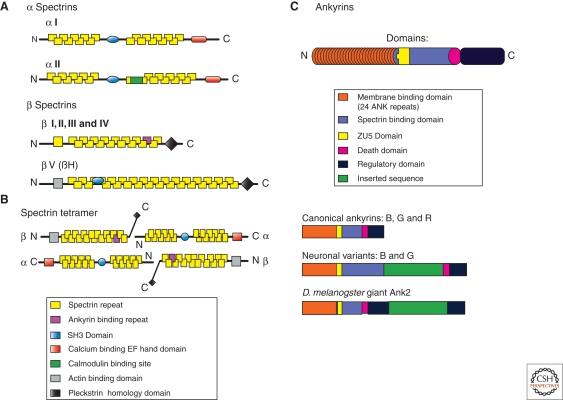
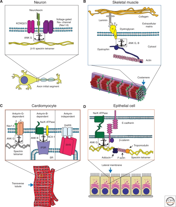
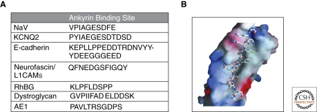
Nodes of Ranvier and axon initial segments of myelinated nerves, sites of cell-cell contact in early embryos and epithelial cells, and neuromuscular junctions of skeletal muscle all perform physiological functions that depend on clustering of functionally related but structurally diverse ion transporters and cell adhesion molecules within microdomains of the plasma membrane. These specialized cell surface domains appeared at different times in metazoan evolution, involve a variety of cell types, and are populated by distinct membrane-spanning proteins. Nevertheless, recent work has shown that these domains all share on their cytoplasmic surfaces a membrane skeleton comprised of members of the ankyrin and spectrin families. This review will summarize basic features of ankyrins and spectrins, and will discuss emerging evidence that these proteins are key players in a conserved mechanism responsible for assembly and maintenance of physiologically important domains on the surfaces of diverse cells.


Figures

Figure 1.
Figure 1.
Domain structure and variants of spectrin and ankyrin proteins. (A) Molecular domains of spectrins: Two α spectrins and five β spectrins are shown. Spectrins are comprised of modular units called spectrin repeats (yellow). Other domains such as the ankyrin binding domain (purple), Src-homology domain 3 (SH3, blue), EF-hand domain (red), and calmodulin-binding domain (green) promote interactions with binding targets important for spectrin function. The pleckstrin homology domain (black) promotes association with the plasma membrane and the actin binding domain (grey) tethers the spectrin-based membrane skeleton to the underlying actin cytoskeleton. (B) The spectrin tetramer, the fundamental unit of the spectrin-based membrane skeleton. The spectrin repeat domains of α and β spectrin associate end-to-end to form heterodimers. Heterodimers associate laterally in an antiparallel fashion to form tetramers. The tetramers can then associate end-to-end to form extended macromolecules that link into a geodesic dome shape directly underneath the plasma membrane. (C) Molecular domains present in canonical ankyrins. The membrane binding domain of ankyrin isoforms (orange) is comprised of 24 ANK repeats. The spectrin binding domain (green-blue) allows ankyrins to coordinate integral membrane proteins to the membrane skeleton. The death domain (pink) is the most highly conserved domain. The regulatory domain (brown) is the most variable region of ankyrins. The regulatory domain interacts intramolecularly with the membrane binding domain to modulate ankyrin’s affinity for other binding partners. All ankyrins and spectrins are subject to alternative splicing, which further increases their functional diversity.
Figure 2.
Figure 2.
Ankyrins and spectrins organize macromolecular complexes in diverse types of specialized membranes. (A) Ankyrin-G forms a complex with β-IV spectrin, neurofascin (a cell adhesion protein), and ion channels (KCNQ2/3 and voltage-gated sodium channel) at axon initial segments in Purkinje neurons. (B) In force buffering costameres of skeletal muscle, ankyrins -B and -G cooperate to target and stabilize key components of the dystroglycoprotein complex. At the membrane, ankyrin-G binds to dystrophin and β-dystroglycan. (C) In cardiomyocyte transverse tubules, ankyrins -B and -G coordinate separate microdomains. Ankyrin-B binds Na+/K+ ATPase, Na+/Ca2+ exchanger (NCX-1), and the inositol triphosphate receptor (IP3R). Ankyrin-G forms a complex with Nav1.5 and spectrin. (D) Ankyrin-G in epithelial lateral membrane assembly. Ankyrin-G binds to E-cadherin, β-2 spectrin, and the Na+/K+ ATPase. Spectrins are connected via F-actin bridges bound to α/γ adducin and tropomodulin.
Figure 3.
Figure 3.
Ankyrins bind to natively unstructured regions of many proteins. (A) Known binding sites of ankyrin proteins. All of these regions lie in regions predicted to be intrinsically unstructured (http://iupred.enzim.hu/IUPs.html). Abbreviations: NaV, voltage-gated sodium channels; KCNQ2, voltage-gated potassium channels; RhBG, rhesus blood group antigen; AE1, anion exchanger. (B) A theoretical model of how the ankyrin membrane binding domain could bind to unstructured peptides. This pocket is 240 angstroms in length with a variety of surface exposed residues.

Similar articles

Cited by

References

    1. Abdi KM, Bennett V 2008. Adducin promotes micrometer-scale organization of β2-spectrin in lateral membranes of bronchial epithelial cells. Mol Biol Cell 19:536–545 - PMC - PubMed
    1. Abdi KM, Mohler PJ, Davis JQ, Bennett V 2006. Isoform specificity of ankyrin-B: A site in the divergent C-terminal domain is required for intramolecular association. J Biol Chem 281:5741–5749 - PubMed
    1. An X, Guo X, Sum H, Morrow J, Gratzer W, Mohandas N 2004. Phosphatidylserine binding sites in erythroid spectrin: Location and implications for membrane stability. Biochemistry 43:310–315 - PubMed
    1. Ango F, di Cristo G, Higashiyama H, Bennett V, Wu P, Huang ZJ 2004. Ankyrin-based subcellular gradient of neurofascin, an immunoglobulin family protein, directs GABAergic innervation at purkinje axon initial segment. Cell 119:257–272 - PubMed
    1. Ayalon G, Davis JQ, Scotland PB, Bennett V 2008. An ankyrin-based mechanism for functional organization of dystrophin and dystroglycan. Cell 135:1189–1200 - PubMed

Publication types

LinkOut - more resources