

The T cells in peripheral taste tissue of healthy human adults: predominant memory T cells and Th-1 cells
- PMID: 20457570
- PMCID: PMC2885746
- DOI: 10.1093/chemse/bjq040
The T cells in peripheral taste tissue of healthy human adults: predominant memory T cells and Th-1 cells
Abstract
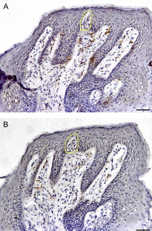
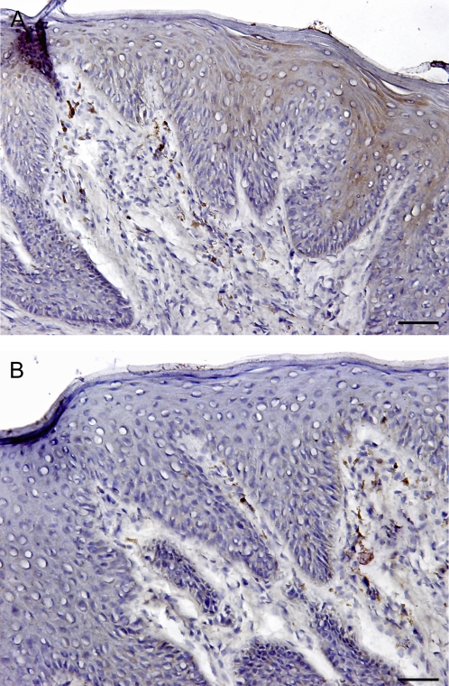
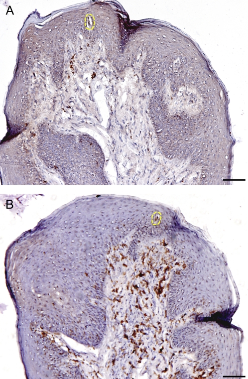
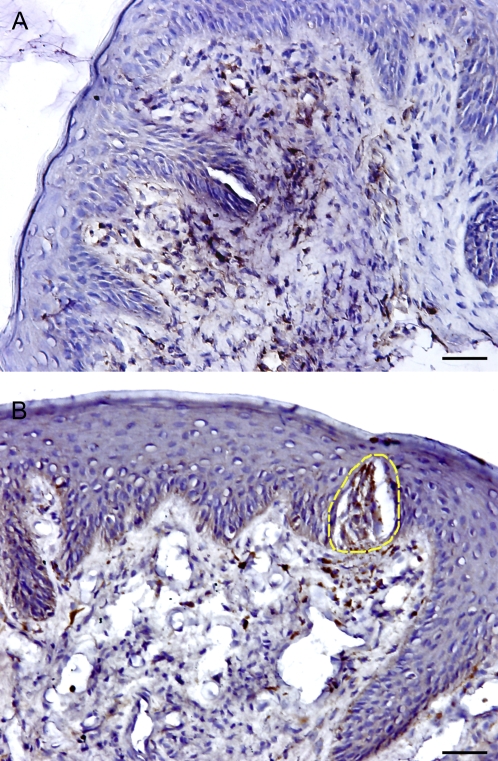
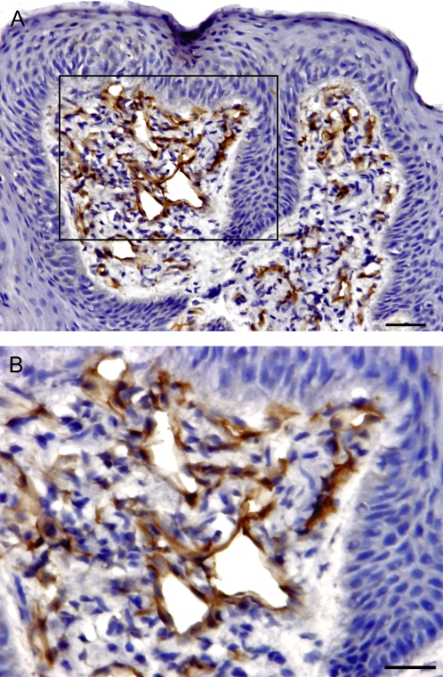
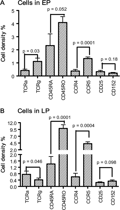
A healthy taste system is important to the maintenance of nutrition and overall quality of life, and taste disorders are associated with many inflammatory states. We previously determined the immune cells in normal human gustatory tissue; they are predominantly dendritic cells and CD4 T cells with a few macrophages and B lymphocytes present. There are, however, few reports of the subtypes of resident lymphocytes in or near taste tissues. The present study further characterized the distribution and population of the major subtypes of T cells in situ within biopsies of healthy human fungiform papillae (FP). Immunohistochemical analyses indicated that T-helper (Th)1 cells (CCR5+) were more predominant in FP than Th2 T cells (CCR4+). CD45RO+ memory T cells were the principal T cells in gustatory tissue, whereas CD45RA+ naive T cells were uncommon. Regarding subcompartments of the tissue, most intraepithelial lymphocytes of FPs were gamma/delta T cells, whereas the major subtype of lymphocytes in the lamina propria were alpha/beta T cells. Regulatory T cells that express CTLA-4 (CD152) and interleukin-2 receptors (IL-2R, CD25) were found at low levels in FP. The T cells stand ready to respond to inflammatory and infectious insults and may play a role in the taste alterations observed during acute and chronic inflammatory states.
Figures
References
-
- Abdollahi M, Radfar M. A review of drug-induced oral reactions. J Contemp Dent Pract. 2003;4(1):10–31. - PubMed
-
- Acuto O, Reinherz EL. The human T-cell receptor. Structure and function. N Engl J Med. 1985;312(17):1100–1111. - PubMed
-
- Baudouin C, Liang H, Bremond-Gignac D, Hamard P, Hreiche R, Creuzot-Garcher C, Warnet JM, Brignole-Baudouin F. CCR 4 and CCR 5 expression in conjunctival specimens as differential markers of T(H)1/ T(H)2 in ocular surface disorders. J Allergy Clin Immunol. 2005;116(3):614–619. - PubMed
-
- Breslin PA, Chapman GB, Mattes RD, Beauchamp GK, Cowart BJ. Quality of life for patients with chemical senses disorders. Chem Senses. 1997;22:650.
-
- Campbell JD, HayGlass KT. T cell chemokine receptor expression in human Th1- and Th2-associated diseases. Arch Immunol Ther Exp (Warsz) 2000;48(6):451–456. - PubMed
Publication types
MeSH terms
Substances
Grants and funding
LinkOut - more resources
Full Text Sources
Research Materials
Miscellaneous
