. 2010 Jul 2;285(27):20564-9.
doi: 10.1074/jbc.M110.132530. Epub 2010 May 10.

The integrator complex recognizes a new double mark on the RNA polymerase II carboxyl-terminal domain

Affiliations

The integrator complex recognizes a new double mark on the RNA polymerase II carboxyl-terminal domain

Sylvain Egloff et al. J Biol Chem. .

Abstract

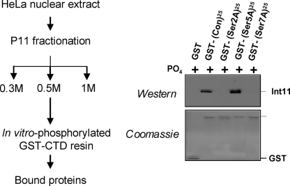
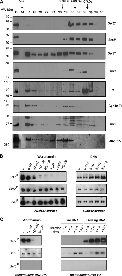
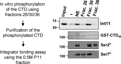
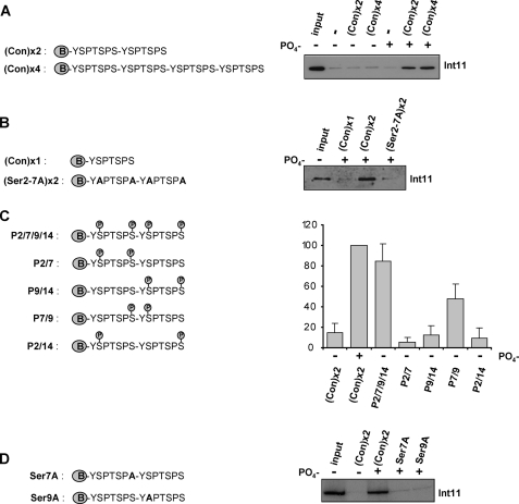
The carboxyl-terminal domain (CTD) of the largest subunit of RNA polymerase II (pol II) comprises multiple tandem repeats of the heptapeptide Tyr(1)-Ser(2)-Pro(3)-Thr(4)-Ser(5)-Pro(6)-Ser(7). This unusual structure serves as a platform for the binding of factors required for expression of pol II-transcribed genes, including the small nuclear RNA (snRNA) gene-specific Integrator complex. The pol II CTD specifically mediates recruitment of Integrator to the promoter of snRNA genes to activate transcription and direct 3' end processing of the transcripts. Phosphorylation of the CTD and a serine in position 7 are necessary for Integrator recruitment. Here, we have further investigated the requirement of the serines in the CTD heptapeptide and their phosphorylation for Integrator binding. We show that both Ser(2) and Ser(7) of the CTD are required and that phosphorylation of these residues is necessary and sufficient for efficient binding. Using synthetic phosphopeptides, we have determined the pattern of the minimal Ser(2)/Ser(7) double phosphorylation mark required for Integrator to interact with the CTD. This novel double phosphorylation mark is a new addition to the functional repertoire of the CTD code and may be a specific signal for snRNA gene expression.


Figures

FIGURE 1.
FIGURE 1.
Both Ser2 and Ser7 of the CTD are required for binding of the Integrator complex. The 0.5 m fraction of P11 fractionation containing the Integrator complex was incubated with in vitro phosphorylated GST-CTD as indicated by the scheme on the left. The panel on the right shows the results of Integrator binding to the wild type CTD (consensus (Con)) and mutant (S2A/S5A/S7A (indicated by Ser2A, Ser5A, and Ser7A)) CTDs assayed by Western blot analysis with antibodies to Int11 (top) and Coomassie Blue staining of the input GST-CTD (bottom).
FIGURE 2.
FIGURE 2.
Ser2 and Ser7 kinase activities can be separated in vitro. A, fractions from HeLa nuclear extract fractionation on a Superose 6 column were tested for the ability to phosphorylate the CTD. The results of Western blot analysis using the phospho-specific antibodies (8) to the modifications noted at the right are shown in the top three panels. The results of Western blot analysis of the fractions using antibodies to the proteins noted at the right are shown in the bottom five panels. The fraction numbers and size of molecular weight (MW) standards are shown at the top. Ip indicates input. Ser2P, Ser(P)2; Ser5P, Ser(P)5; Ser7P, Ser(P)7. B, Western blot analysis of GST-CTD phosphorylation by nuclear extract supplemented with wortmannin (left panel) or DNA (right panel) using the phospho-specific antibodies noted at the left. C, Western blot analysis of GST-CTD phosphorylation by recombinant DNA-PK supplemented with wortmannin (left panel) or DNA (as a time course) (right panel) using the phospho-specific antibodies noted at the left.
FIGURE 3.
FIGURE 3.
Phosphorylation of both Ser2 and Ser7 is required for Integrator binding. Fractions (Frac.) containing Ser2 kinase activity, Ser7 kinase activity, or both were used to phosphorylate GST-CTD and tested for their ability to bind the Integrator complex, as indicated in the scheme on the left. The results of Western blot analysis using anti-Int11 antibodies (23) (top), Coomassie Blue staining of the input GST-CTD (middle), and Western blot analysis using antibodies to phospho-Ser2 (Ser2P) and phospho-Ser7 (Ser7P) (bottom) are shown on the right. NE = nuclear extract.
FIGURE 4.
FIGURE 4.
A specific double mark on the CTD directs the binding of Integrator. A–D, the structures of the biotinylated peptides used are shown on the left. The results of Western blot analysis using anti-Int11 (23) of proteins pulled down by the peptides noted above the lanes are shown on the right. In panel C, quantitation is shown underneath with error bars indicating the S.D. from four independent experiments. Con, consensus.

References

    1. Hirose Y., Ohkuma Y. (2007) J. Biochem. 141, 601–608 - PubMed
    1. Chapman R. D., Heidemann M., Hintermair C., Eick D. (2008) Trends Genet. 24, 289–296 - PubMed
    1. Egloff S., Murphy S. (2008) Trends Genet. 24, 280–288 - PubMed
    1. Buratowski S. (2003) Nat. Struct. Biol. 10, 679–680 - PubMed
    1. Corden J. L. (2007) Science 318, 1735–1736 - PubMed

Publication types

MeSH terms