. 2010 Aug 13;285(33):25285-95.
doi: 10.1074/jbc.M109.087239. Epub 2010 May 10.

CIN85/RukL is a novel binding partner of nephrin and podocin and mediates slit diaphragm turnover in podocytes

Affiliations

CIN85/RukL is a novel binding partner of nephrin and podocin and mediates slit diaphragm turnover in podocytes

Irini Tossidou et al. J Biol Chem. .

Abstract

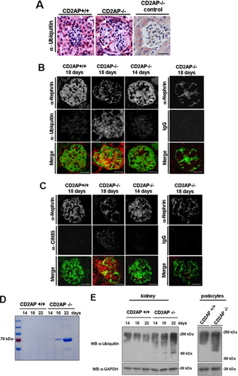
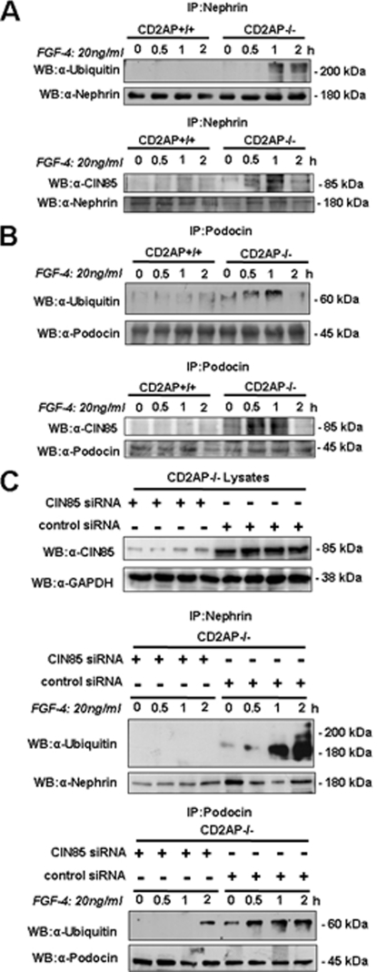
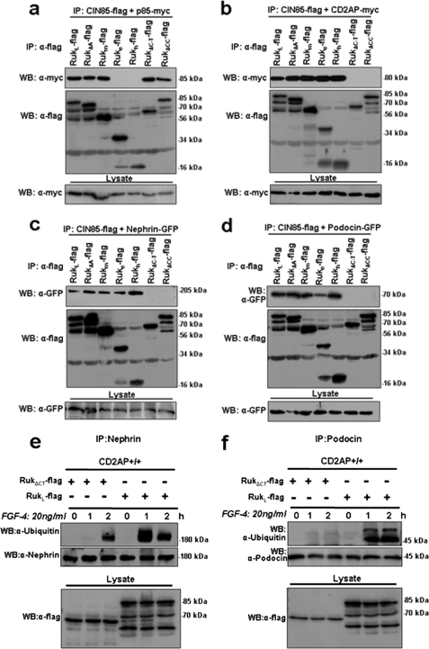
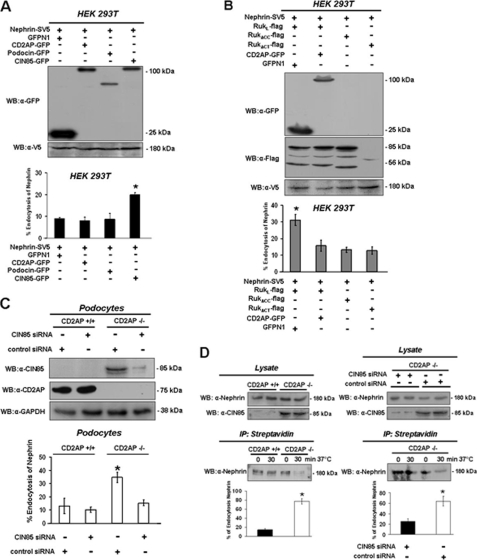
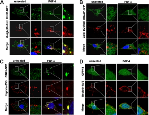
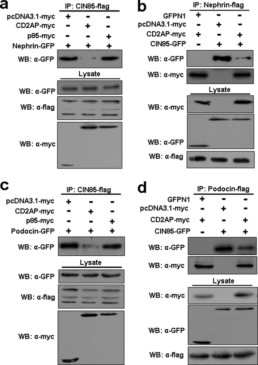
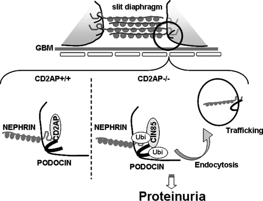
Podocyte damage is the basis of many glomerular diseases with ultrastructural changes and decreased expression of components of the slit diaphragm such as nephrin and podocin. Under physiological conditions it is likely that the slit diaphragm underlies permanent renewal processes to indemnify its stability in response to changes in filtration pressure. This would require constant reorganization of the podocyte foot process and the renewal of slit diaphragm components. Thus far, the mechanisms underlying the turnover of slit diaphragm proteins are largely unknown. In this manuscript we examined a mechanism of nephrin endocytosis via CIN85/Ruk(L)-mediated ubiquitination. We can demonstrate that the loss of nephrin expression and onset of the proteinuria in CD2AP(-/-) mice correlates with an increased accumulation of ubiquitinated proteins and expression of CIN85/Ruk(L) in podocytes. In cultured murine podocytes CD2AP deficiency leads to an early ubiquitination of nephrin and podocin after stimulation with fibroblast growth factor-4. Binding assays with different CIN85/Ruk isoforms and mutants showed that nephrin and podocin are binding to the coiled-coil domain of CIN85/Ruk(L). We found that in the presence of CIN85/Ruk(L), which is involved in down-regulation of receptor-tyrosine kinases, nephrin is internalized after stimulation with fibroblast growth factor-4. Interestingly, coexpression of CIN85/Ruk(L) with CD2AP led to a decreased binding of CIN85/Ruk(L) to nephrin and podocin, which indicates a functional competition between CD2AP and CIN85/Ruk(L). Our results support a novel role for CIN85/Ruk(L) in slit diaphragm turnover and proteinuria.


Figures

FIGURE 1.
FIGURE 1.
Onset of proteinuria correlates with accumulation of ubiquitinated proteins in podocytes of CD2AP−/− mice. A, immunohistochemistry using an anti-ubiquitin antibody shows ubiquitin-positive podocytes of diseased 3-week-old CD2AP−/− mice (white arrows). As a control the primary antibody was replaced by rabbit IgG. B and C, fluorescence labeling demonstrates ubiquitin (B) or CIN85/RukL (C) (red fluorescence) and nephrin (green fluorescence) in renal cortex sections of 18-day-old CD2AP+/+ and proteinuric CD2AP−/− mice as well as healthy 14-day-old CD2AP−/− mice as control. Glomerular ubiquitin-positive cells and CIN85/RukL expression is increased, whereas nephrin expression is down-regulated in 18-day-old CD2AP−/− mice. Ubiquitin and CIN85/Ruk partially colocalize with nephrin resulting in yellow fluorescence in the merged picture. Control staining was performed with mouse or rabbit IgG instead of the primary antibody. The size of the scale bars is 30 μm. D, spot urine of 14-, 18-, and 22-day-old CD2AP+/+ and CD2AP−/− mice was used for SDS-PAGE and stained with Coomassie Blue. The onset of proteinuria is clearly detectable at 18 days in the CD2AP−/− mice. E, whole kidney lysates of 14-, 18-, and 22-day-old CD2AP+/+ and CD2AP−/− mice (left) and protein lysates from cultured CD2AP+/+ and CD2AP−/− podocytes (right) were analyzed for ubiquitin by Western blot (WB). CD2AP−/− kidneys and cultured podocytes show an accumulation of ubiquitinated proteins. GAPDH, glyceraldehyde-3-phosphate dehydrogenase.
FIGURE 2.
FIGURE 2.
Nephrin and podocin are early ubiquitinated in CD2AP−/− podocytes after growth factor stimulation. CD2AP+/+ and CD2AP−/− podocytes were stimulated for 0, 0.5, 1, and 2 h with FGF-4, and the whole cell lysate was then precipitated (IP) with nephrin (A) or podocin (B). The probes were blotted and analyzed for ubiquitin, CIN85/RukL, nephrin, and podocin. C, CD2AP−/− podocytes were treated with 20 pmol of CIN85/RukL siRNA or control siRNA (scrambled sequence) and cultured for 72 h. Lysates were then analyzed for CIN85/RukL and glyceraldehyde-3-phosphate dehydrogenase (GAPDH) to show inhibited expression of CIN85/RukL (upper panel). Silenced cells were stimulated with FGF-4 for 0, 0.5, 1, and 2 h, and the whole cell lysates were then precipitated with anti-nephrin (middle) and anti-podocin (lower panels). Precipitates where then analyzed for ubiquitin content. WB, Western blot.
FIGURE 3.
FIGURE 3.
CIN85/RukL interacts with nephrin and podocin at the coiled-coil domain. HEK 293T cells were transiently transfected with constructs encoding FLAG-tagged isoforms of CIN85/Ruk and Nephrin-GFP, Podocin-GFP, CD2AP-myc, or p85-myc. CIN85/Ruk-FLAG was precipitated (IP) from whole cell lysate using FLAG beads. The probes were blotted (WB) and analyzed for GFP or myc expression. For control of expression whole cell lysates were blotted and analyzed for FLAG, GFP, or myc expression. The binding of p85 (a) and CD2AP (b) to the Ruk isoforms was performed as a control for specific binding and proof of integrity of the coiled-coil domain. Nephrin (c) and podocin (d) interact with the coiled-coil domain of CIN85/Ruk. CD2AP+/+ podocytes were transiently transfected with FLAG-RukLΔCT or FLAG-RukL and stimulated for 0, 1, and 2 h with FGF-4, and the whole cell lysates were precipitated with anti-nephrin (e) or anti-podocin (f). The probes were then blotted and analyzed for ubiquitin, nephrin or podocin. Lysates were analyzed for FLAG expression.
FIGURE 4.
FIGURE 4.
Endocytosis of nephrin is enhanced in the presence of CIN85/RukL. HEK 293T cells were cotransfected with Nephrin-SV5 and GFPN1, CD2AP-GFP, Podocin-GFP, or CIN85-GFP (A) or with Nephrin-SV5 and GFPN1, RukL,-FLAG, RukL-FLAG with CD2AP-GFP, RukΔCC-FLAG, or RukΔCT-FLAG (B). WB, Western blot. Nephrin-SV5 expressed at the surface of the cells was labeled with an anti-V5-antibody. The cells were labeled at 4 °C followed by incubation at 37 °C for 60 min to induce endocytosis. The controls remained at 4 °C. The extinction was measured at 405 nm. A, *, p < 0.007 CIN85/RukL versus GFPN1, CD2AP, and Podocin. B, *, p < 0.04 RukL versus Rukl + CD2AP, RukΔCC, and RukΔCT. The upper panels show expression of proteins analyzed by Western blot. C, CD2AP+/+ and CD2AP−/− were treated with a CIN85/RukL siRNA or control siRNA and cultured for 72 h. Surface-expressed nephrin was labeled with an anti-nephrin antibody. Endocytosis of endogenous nephrin was measured by the same method. *. p < 0.05, CD2AP−/− podocytes treated with control siRNA versus CD2AP−/− podocytes treated with CIN85/RukL siRNA and CD2AP+/+ podocytes treated with control siRNA. The upper panels show expression of proteins analyzed by Western blot. GAPDH, glyceraldehyde-3-phosphate dehydrogenase. D, podocyte surface proteins were biotinylated in CD2AP+/+ and CD2AP−/− podocytes, and CD2AP−/− podocytes were treated with a CIN85/RukL siRNA and control siRNA. Lysates were precipitated (IP) using streptavidin-coupled beads, and nephrin expression was analyzed using a Western blot. Left panel, *, p < 0.005, CD2AP−/− versus CD2AP+/+ podocytes; right panel, *, p < 0.04 CD2AP−/− podocytes treated with control siRNA versus CD2AP−/− podocytes treated with CIN85/RukL siRNA. The upper panels show expression of nephrin and CIN85/RukL in the lysates.
FIGURE 5.
FIGURE 5.
Nephrin is internalized after growth factor stimulation in the presence of CIN85/RukL. A and B, CD2AP+/+ podocytes were co-transfected with CIN85-GFP (A) or CD2AP-GFP (B) and β-1,4-galactotransferase-pDsRed (Golgi-associated protein). The cells were left untreated or stimulated for 60 min with FGF-4 (20 ng/ml). CIN85/RukL (green) colocalizes with β-1, 4-galactotransferase (red) after treatment with FGF-4. C and D, CD2AP+/+ podocytes were co-transfected with Nephrin-SV5 and with CIN85-GFP or GFPN1 empty vector. Nephrin was visualized by staining the living cells with an anti-SV5 antibody and a Cy3-labeled secondary antibody. The cells were left untreated or stimulated for 60 min with FGF-4 (20 ng/ml). Nephrin (red) is expressed alongside the cell membrane in unstimulated cells. After stimulation, nephrin (red) is distributed perinuclear in the presence of CIN85/RukL (green). Colocalization results in yellow fluorescence in the merged pictures. Nuclei were stained with 4′,6-diamidino-2-phenylindole for visualization. Size of scale bars is 10 μm.
FIGURE 6.
FIGURE 6.
Nephrin and podocin bind preferentially to CD2AP. HEK 293T cells were transiently transfected with CD2AP, CIN85/RukL, nephrin, and podocin. CIN85/RukL-FLAG, Nephrin-FLAG, or Podocin-FLAG was precipitated (IP) from whole cell lysates using anti-FLAG beads. The probes were analyzed for GFP or myc expression. Lysates were blotted (WB) and analyzed for protein content using anti-GFP, FLAG, or myc antibodies. In the presence of CD2AP, binding of CIN85/RukL to nephrin is significantly reduced (a and b). Similarly, the binding of podocin to CIN85/RukL is significantly impaired in the presence of CD2AP (c and d).
FIGURE 7.
FIGURE 7.
Working model of slit diaphragm turnover and trafficking of nephrin and podocin. In the presence of CD2AP the slit diaphragm complexes are stabilized. If CD2AP dissociates or is absent, CIN85 can bind to both molecules and induce trafficking of nephrin and podocin. Ubi, ubiquitin; GBM, glomerular basement membrane.

Similar articles

Cited by

References

    1. Kerjaschki D. (2001) J. Clin. Invest. 108, 1583–1587 - PMC - PubMed
    1. Tryggvason K., Wartiovaara J. (2001) Curr. Opin. Nephrol. Hypertens. 10, 543–549 - PubMed
    1. Huber T. B., Kottgen M., Schilling B., Walz G., Benzing T. (2001) J. Biol. Chem. 276, 41543–41546 - PubMed
    1. Huber T. B., Hartleben B., Kim J., Schmidts M., Schermer B., Keil A., Egger L., Lecha R. L., Borner C., Pavenstädt H., Shaw A. S., Walz G., Benzing T. (2003) Mol. Cell. Biol. 23, 4917–4928 - PMC - PubMed
    1. Schiffer M., Mundel P., Shaw A. S., Bottinger E. P. (2004) J. Biol. Chem. 279, 37004–37012 - PubMed

Publication types

MeSH terms

LinkOut - more resources