. 2010 Jul 16;285(29):22003-16.
doi: 10.1074/jbc.M110.101923. Epub 2010 May 10.

The KRAS promoter responds to Myc-associated zinc finger and poly(ADP-ribose) polymerase 1 proteins, which recognize a critical quadruplex-forming GA-element

Affiliations

The KRAS promoter responds to Myc-associated zinc finger and poly(ADP-ribose) polymerase 1 proteins, which recognize a critical quadruplex-forming GA-element

Susanna Cogoi et al. J Biol Chem. .

Abstract

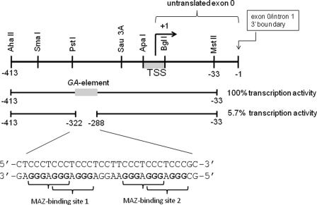
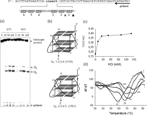
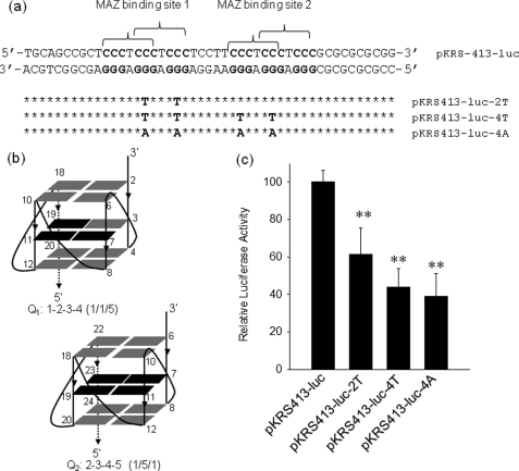
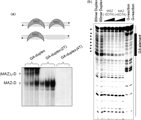
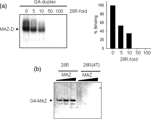
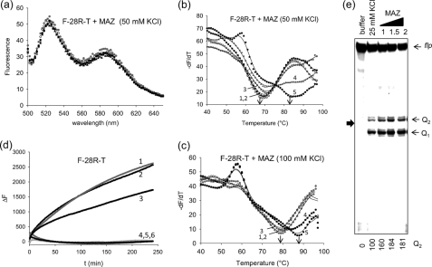
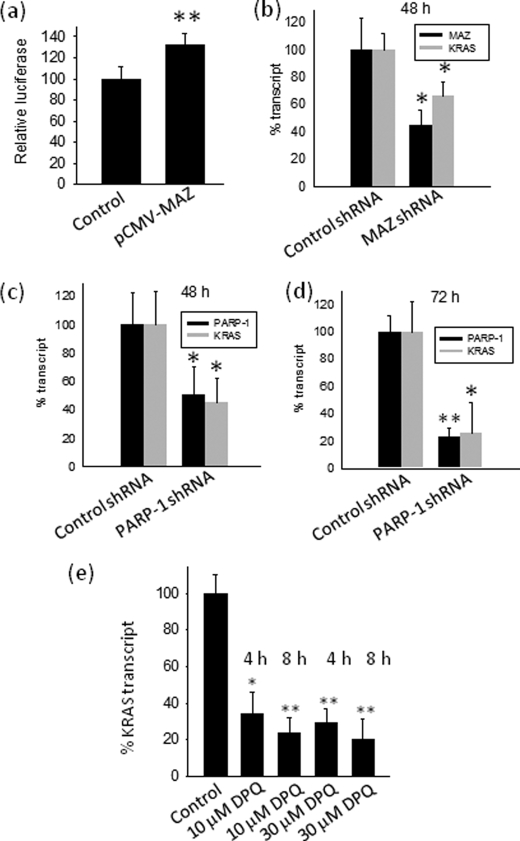
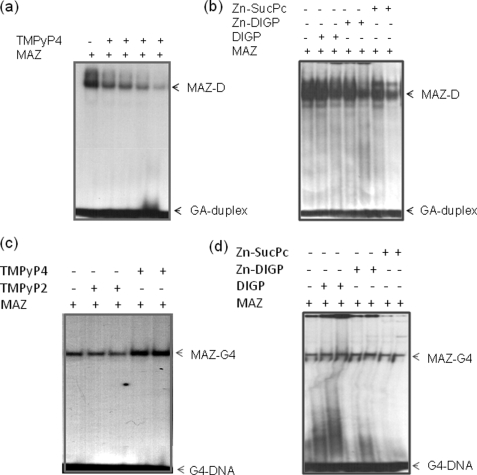
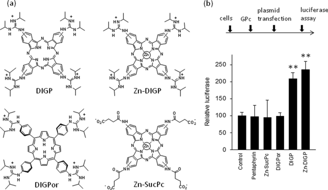
The murine KRAS promoter contains a G-rich nuclease hypersensitive element (GA-element) upstream of the transcription start site that is essential for transcription. Pulldown and chromatin immunoprecipitation assays demonstrate that this GA-element is bound by the Myc-associated zinc finger (MAZ) and poly(ADP-ribose) polymerase 1 (PARP-1) proteins. These proteins are crucial for transcription, because when they are knocked down by short hairpin RNA, transcription is down-regulated. This is also the case when the poly(ADP-ribosyl)ation activity of PARP-1 is inhibited by 3,4-dihydro-5-[4-(1-piperidinyl) butoxyl]-1(2H) isoquinolinone. We found that MAZ specifically binds to the duplex and quadruplex conformations of the GA-element, whereas PARP-1 shows specificity only for the G-quadruplex. On the basis of fluorescence resonance energy transfer melting and polymerase stop assays we saw that MAZ stabilizes the KRAS quadruplex. When the capacity of folding in the GA-element is abrogated by specific G --> T or G --> A point mutations, KRAS transcription is down-regulated. Conversely, guanidine-modified phthalocyanines, which specifically interact with and stabilize the KRAS G-quadruplex, push the promoter activity up to more than double. Collectively, our data support a transcription mechanism for murine KRAS that involves MAZ, PARP-1 and duplex-quadruplex conformational changes in the promoter GA-element.


Figures

FIGURE 1.
FIGURE 1.
The murine KRAS promoter contains a nuclease hypersensitive G-rich element (GA-element) that is essential for transcription. The GA-element is characterized by six runs of guanines that can fold into an intramolecular G-quadruplex structure. The GA-element contains two perfect binding sites for the MAZ transcription factor. TSS means transcription start site. The sequence is numbered with the exon 0/intron 1 boundary taken as −1.
FIGURE 2.
FIGURE 2.
Polymerase stop and FRET-melting assays. The DNA template used for the polymerase stop assay is shown. a, the G-runs of the GA-element are underlined. Primer-extension reactions were performed at 37 and 50 °C. Two polymerase pauses were observed at the adenines (determined by Sanger sequencing, see Ref. and supplemental Fig. S1) before the first and second G-run at the 3′ end of the GA-element. The numbers above the gel indicate the KCl concentrations in the samples. b, shown are structures of the putative G-quadruplexes determined by dimethyl sulfate footprinting and circular dichroism (see Ref. 7). c, energy transfer, p = I580/(I525 + I580)-associated to the folding of F-28R-T as a function of KCl concentration, is shown. The experiment was performed in 50 mm Tris-HCl, pH 7.4, KCl as indicated in the figure (10, 25, 50, 100, and 140 mm). d, shown are FRET-melting curves obtained by measuring the increase of fluorescence at 525 nm emitted by F-28R-T as a function of temperature (excitation, 485 nm). The numbers indicate the KCl concentration. F-28R-T melts with a biphasic profile in the presence of 100 and 140 mm KCl.
FIGURE 3.
FIGURE 3.
Transcription activity of wild-type and mutant KRAS promoter. a, shown is the sequence of the KRAS promoter in the region spanning over the GA-element. The two MAZ-binding sites are indicated by brackets. By site-directed mutagenesis the promoter sequence was modified in the GA-element either in one MAZ-binding site or in both MAZ-binding sites. The point mutations abrogated the capacity of the sequence to fold into a G-quadruplex. b, the structure of the putative KRAS G-quadruplexes shows that the point mutations fall in the mid G-tetrad of the structure (depicted in black), c, results of dual luciferase assay with wild-type and mutant plasmids show that the activity of the KRAS promoter is reduced by the introduction in the GA-element of the point mutations that destabilize G-quadruplex formation. All mutant expressions are different than wild-type expression by Student's t test; p < 0,01 (two asterisks).
FIGURE 4.
FIGURE 4.
Binding of MAZ to duplex of the GA-element. a, an EMSA shows the formation of two DNA·MAZ complexes with a 1:1 and 1:2 stoichiometry. The targets used are the GA-duplex, the mutant duplexes with 2 and 4 G → T mutations (GA-duplex (2T) and GA-duplex (4T) were obtained annealing 28R(2T) and 28R(4T) (Table 1) to their complementary oligonucleotides). MAZ amounts of 1, 2, and 4 μg were used, whereas the target 32P duplex was 20 nm. The experiment was performed with 5% PAGE in TBE. b, DNase I footprinting of an 80-mer promoter fragment containing the GA-element is shown. From left, first and second lanes, duplex digested with DNase I; third-fifth lanes and sixth-eighth lanes, DNase I digestion in the absence and presence of EDTA in the presence of 0.5, 1 and 2 μm MAZ-GST.
FIGURE 5.
FIGURE 5.
EMSA competition experiments and binding of MAZ to G4-DNA. a, a competition assay shows that the DNA-MAZ complex is competed away by 5-, 10-, 50-, and 100-fold of cold quadruplex 28R. Experiments performed with 20 nm GA-duplex and 3 μg of MAZ-GST. Histograms show the densitometric analysis of the gel. b, an EMSA shows the binding of quadruplex 28R to the MAZ protein. Radiolabeled quadruplex 28R and mutant 28R(4T) (20 nm) have been incubated with 2, 3, and 5 μg MAZ. 5% PAGE in TBE.
FIGURE 6.
FIGURE 6.
MAZ stabilizes the G-quadruplex. a, emission spectra are shown of quadruplex F-28R-T treated with BSA (r ([protein])/[quadruplex]) = 5) or increasing amounts of recombinant MAZ (r = 0, 1, 2. 5, 5). Excitation 475 nm. The experiment was carried out in 50 mm Tris-HCl, pH 7.4, 50 mm KCl, 50 μm zinc acetate; spectra were recorded using a fluorometer. b, shown is FRET melting of the same samples described in a. Curves 1 and 2, F-28R-T with and without BSA; curves 3, 4, and 5, F-28R-T with MAZ at r = 1, 2.5, and 5; the experiment was carried out in 50 mm Tris-HCl, pH 7.4, 50 mm KCl, 50 μm zinc acetate. c, the experiment was as in b but with 100 mm KCl. d, shown is the rate of assembly between quadruplex F-28R-T and 28Y. Curve 1, F-28R-T + 28Y (1:1); curve 2, F-28R-T + 28Y (1:1) + BSA (r = 2); curve 3, F-28R-T + 28Y (1:1) + MAZ (r = 1); curve 4, F-28R-T + 28Y (1:1) + MAZ (r = 2); curve 5, F-28R-T + BSA (r = 2); curve 6, F-28R-T MAZ (r = 2). The experiment was carried out in 50 mm Tris-HCl, pH 7.4, 50 mm KCl, 50 μm zinc acetate at 37 °C. The data shown in panels b–d have been collected by a real time apparatus (CFX96 Bio-Rad) measuring the FAM emission at 525 nm. e, shown is a polymerase-stop assay using the wtR-Mur80 template, 18-mer primer pMur80, and increasing amounts of MAZ (1, 1.5, and 2 μg) as indicated in the figure. The experiment was carried out in 50 mm Tris-HCl, pH 7.4, 25 mm KCl, 50 μm zinc acetate, 12% polyacrylamide gel in 1× TBE. flp, full-length product. Numbers at the bottom indicate the intensity of the Q2 band.
FIGURE 7.
FIGURE 7.
Pulldown and ChIP assays. a, biotinylated GA-element in duplex (G4-biotin hybridized to its complementary) or in quadruplex (G4-biotin) were used as bait in pulldown experiments with NIH 3T3 extract. The concentrations of NaCl in the elution buffer used to elute the protein fractions were 0.5 and 1 m. The panels show the Western blots of the pulled down fractions obtained with anti MAZ (top panel) and anti-PARP-1 (bottom panel) antibodies. The band intensities have been measured with ChemiDOC XRS apparatus (Bio-Rad). b, a chromatin immunoprecipitation assay was performed with anti-MAZ, anti-PARP-1, anti- RNA polymerase II (positive control), and IgG (negative control) antibodies. PCR analysis was performed on DNA isolated from ChIP reactions using controls, anti-PARP-1, and anti MAZ antibodies. PCR was performed with KRAS primers (see “Experimental Procedures”) and EF1-α control primers (EF1-α primers provided by the kit amplify a 233-bp fragment from the DNA immunoprecipitated with anti-RNA polymerase II, used as a positive control). The KRAS PCR amplification product obtained with anti MAZ and anti-PARP-1antibodies show that under in vivo conditions the GA-element is bound by PARP-1 and MAZ.
FIGURE 8.
FIGURE 8.
Effect of MAZ and PARP-1 on transcription. a, transient transfection experiments show the effect of MAZ overexpression on firefly luciferase driven by the wild-type KRAS promoter. Left bar (control), cells transfected with pKRS413-luc, pcDNA3 (empty vector), and pRL-CMV; right bar, cells transfected with pKRS413-luc, pCMV-MAZ, and pRL-CMV. The ordinate reports firefly luciferase normalized by Renilla luciferase; b–d, real-time PCR shows the effect on KRAS transcription of knocking down MAZ or PARP-1 with specific shRNAs at 48 and 72 h. The ordinate reports the % transcript (KRAS or MAZ or PARP-1), i.e. RT/RC × 100, where RT is (transcript)/(average transcripts from β2-microglobulin, glyceraldehyde-3-phosphate dehydrogenase, and hypoxanthine-guanine phosphoribosyltransferase) in shRNA-treated cells, and RC is (transcript)/(average transcripts from β2-microglobulin, glyceraldehyde-3-phosphate dehydrogenase, and hypoxanthine-guanine phosphoribosyltransferase) in cells treated with unrelated shRNA. e, real time PCR shows the effect on KRAS transcription of 10 and 30 μm DPQ, an inhibitor of PARP-1 poly (ADP-ribose) activity. The ordinate reports the % KRAS transcript, i.e. RT/RC × 100, where RT is (KRAS transcript)/(β2microglobulin and hypoxanthine-guanine phosphoribosyltransferase transcripts) in untreated cells, and RC is (KRAS transcript)/(β2-microglobulin and hypoxanthine-guanine phosphoribosyltransferase transcripts) in cells treated with DPQ. Differences from the control are supported by Student's t test, p < 0.05 (one asterisk), p < 0.01 (two asterisks).
FIGURE 9.
FIGURE 9.
Effect of G4-DNA ligands on KRAS transcription. a, an EMSA shows that the complex between GA-duplex and MAZ-GST is competed by increasing amounts of TMPyP4 (0.5, 1, 2, and 5 μm). Duplex concentration is 20 nm, and MAZ is 4 μg (5% PAGE in TBE). b, shown is the same experiment as in a but with phthalocyanines DIGP, Zn-DIGP, and Zn-SucPc at concentrations 5 and 10 μm. c, an EMSA show that the KRAS G-quadruplex binds to MAZ-GST in the absence and presence of TMPyP2 or TMPyP4 (2.5 and 10 μm), 10% PAGE in TBE. d, shown is the same experiment as in c but phthalocyanines DIGP, Zn-DIGP, and Zn-SucPc at the concentrations of 5 and 10 μm. The streaking in the second, third, and fifth lanes from the left is due to the effect of phthalocyanines binding to the quadruplex.
FIGURE 10.
FIGURE 10.
Transcription assays. a, structures of guanidine phthalocyanines used are shown. b, a dual luciferase assay was performed in NIH 3T3 cells treated with 10 μm DIGP, Zn-DIGP, Zn-SucPc, DIGPor, or pentaphyrin for 16 h and subsequently transfected with a mixture of pKRS413-luc and pRL-CMV. The signal was normalized to Renilla luciferase (control). Luciferase expressions in the presence of DIGP and Zn-DIGP are different from control, by Student's t test, p < 0.01 (two asterisks).

Similar articles

Cited by

References

    1. Sen D., Gilbert W. (1988) Nature 334, 364–366 - PubMed
    1. Maizels N. (2006) Nat. Struct. Mol. Biol. 13, 1055–1059 - PubMed
    1. Huppert J. L., Balasubramanian S. (2007) Nucleic Acids Res. 35, 406–413 - PMC - PubMed
    1. Todd A. K., Johnston M., Neidle S. (2005) Nucleic Acids Res. 33, 2901–2907 - PMC - PubMed
    1. Eddy J., Maizels N. (2006) Nucleic Acids Res. 34, 3887–3896 - PMC - PubMed

Publication types

MeSH terms