. 2010 Jul 16;285(29):22202-10.
doi: 10.1074/jbc.M110.131821. Epub 2010 May 10.

The anti-inflammatory prostaglandin 15-deoxy-delta(12,14)-PGJ2 inhibits CRM1-dependent nuclear protein export

Affiliations

The anti-inflammatory prostaglandin 15-deoxy-delta(12,14)-PGJ2 inhibits CRM1-dependent nuclear protein export

Mark Hilliard et al. J Biol Chem. .

Abstract

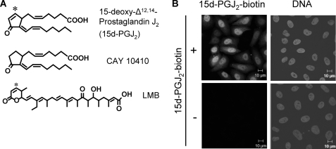
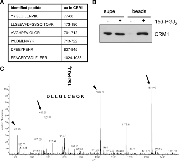
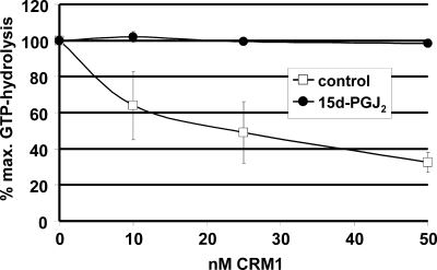
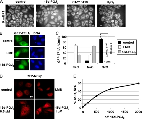
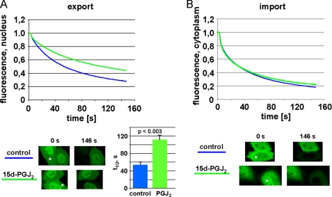
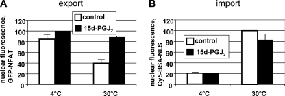
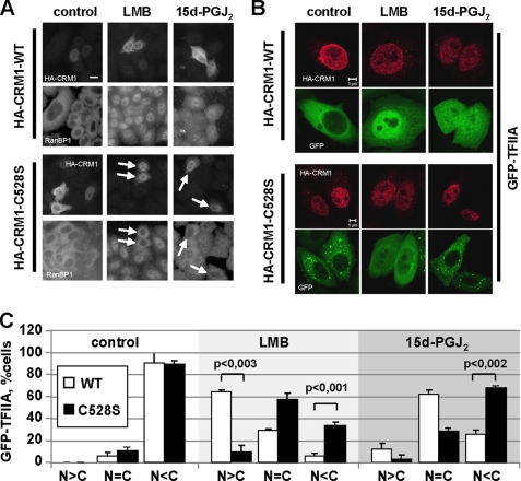
The signaling molecule 15-deoxy-Delta(12,14)-prostaglandin J(2) (15d-PGJ(2)) has been described as the "anti-inflammatory prostaglandin." Here we show that substrates of the nuclear export receptor CRM1 accumulate in the nucleus in the presence of 15d-PGJ(2), identifying this prostaglandin as a regulator of CRM1-dependent nuclear protein export that can be produced endogenously. Like leptomycin B (LMB), an established fungal CRM1-inhibitor, 15d-PGJ(2) reacts with a conserved cysteine residue in the CRM1 sequence. This covalent modification prevents the formation of nuclear export complexes. Cells that are transfected with mutant CRM1 (C528S) are resistant to the inhibitory effects of LMB and 15d-PGJ(2), demonstrating that the same single amino acid is targeted by the two compounds. Inhibition of the CRM1 pathway by endogenously produced prostaglandin and/or exogenously applied 15d-PGJ(2) may contribute to its anti-inflammatory, anti-proliferative, and anti-viral effects.


Figures

FIGURE 1.
FIGURE 1.
15d-PGJ2 accumulates in nuclei. A, chemical structures of 15d-PGJ2, CAY 10410 (9,10-dihydro-15-deoxy-Δ12,14-prostaglandin J2) and LMB. The asterisks indicate the reactive C-atoms in 15d-PGJ2 and LMB. B, HeLa-cells were treated with 10 μm 15d-PGJ2-biotin for 1 h, fixed, and stained with Alexa488-streptavidin (left) and Hoechst (DNA, right).
FIGURE 2.
FIGURE 2.
CRM1 is a target of 15d-PGJ2. A, identified CRM1-peptides. Biotin-15d-PGJ2-modified proteins from a HeLa-cell lysate were subjected to LC-ESI-MS/MS. The identity of peptide LLSEEVFDFSSGQITQVK was confirmed by sequencing (compare supplemental Fig. S1). The amino acids correspond to the human CRM1-sequence. B, a cell lysate was treated with (+) or without (−) biotinylated 15d-PGJ2 and neutravidin-binding proteins (beads) were analyzed by Western blotting using an anti-CRM1 antibody. 1% of the supernatant (supe) was loaded. C, full mass spectrum of a CRM1 peptide modified by 15d-PGJ2. The peptide DLLGLCEQK (amino acids sequence 523–531 of CRM1) was treated with 15d-PGJ2, and the samples were analyzed by mass spectrometry. The arrows show the peaks 667 and 1334, corresponding to the modified peptide doubly and singly charged, respectively. The arrowheads show the peaks corresponding to the unmodified peptides.
FIGURE 3.
FIGURE 3.
15d-PGJ2 inhibits the formation of trimeric export complexes. RanGAP assays were performed in the presence of an NES-peptide and increasing concentrations of CRM1 that had been preincubated with 15d-PGJ2 (closed circles) or ethanol as a control (open squares). Trimeric export complexes are insensitive to GTPase activation on Ran by RanGAP. High levels of GTP hydrolysis indicate the presence of free RanGTP in the reaction. Bars indicate the variation from the mean of two independent experiments.
FIGURE 4.
FIGURE 4.
15d-PGJ2 inhibits nuclear export in vivo. A, 15d-PGJ2 inhibits nuclear export of RanBP1. Cells were treated with 30 μm of 15d-PGJ2 or CAY10410 or 10 mm H2O2 for 1.5 h as indicated and analyzed for localization of endogenous RanBP1. B and C, cells were transfected with a plasmid coding for GFP-TFIIA and treated with ethanol as a control or 5 nm LMB or 30 μm 15d-PGJ2, as indicated. C, quantification of the subcellular localization of GFP-TFIIA. Whereas ∼95% of all cells showed a more cytoplasmic localization (N<C) of GFP-TFIIA under control conditions, this percentage significantly decreased in the presence of either LMB or 15d-PGJ2. D, HeLa cells were transfected with a plasmid coding for RFP-NC2β and treated for 2 h with ethanol (control), 5 nm LMB, or 0.5 or 1 μm 15d-PGJ2 as indicated. E, quantification of cells showing a clear nuclear localization of RFP-NC2β (N>C) in the presence of increasing concentrations of 15d-PGJ2. Error bars in C and E (sometimes too small to be seen) indicate the standard deviation from the mean of three independent experiments, counting ∼100 cells per condition. p values in E (in relation to 0 nm) were < 0.01 for concentrations of 15d-PGJ2 up to 500 nm and <0.001 for higher concentrations. Bars, 10 μm (A, B, D).
FIGURE 5.
FIGURE 5.
FLIP analysis of export inhibition by 15d-PGJ2. Cells were transfected with a plasmid coding for NES-GFP2-M9 and subjected to FLIP-analysis in the presence of 15 μm 15d-PGJ2 or ethanol as a control. The graphs show the mean of three (A, export) or two (B, import) independent experiments, analyzing 10–20 cells each. Error bars are omitted for clarity (see supplemental Fig. S3 for the original data set of a single experiment). A, a region in the cytoplasm was bleached to analyze loss of fluorescence from the nucleus, i.e. nuclear export. The bar graph shows t½, i.e. the time it takes to loose 50% of the nuclear fluorescence. The bars indicate the standard deviation from the mean of three independent experiments. B, a region in the nucleus was bleached to analyze loss of fluorescence from the cytoplasm, i.e. nuclear import. Single cells before the first (t = 0 s) and after the last (t = 146 s) bleach interval are depicted. Asterisks indicate the bleached compartments. Note that in the ethanol control (A) the bleached cell looses the vast majority of its fluorescence, indicating efficient export. The 15d-PGJ2-treated cell, by contrast, retains a significant level of fluorescence in the nucleus, indicating inhibited export.
FIGURE 6.
FIGURE 6.
15d-PGJ2 inhibits nuclear export, but not import, in vitro. After digitonin permeabilization and preincubation with 15d-PGJ2 (black bars) or ethanol as a control (gray bars), GFP-NFAT-expressing cells were subjected to nuclear transport reactions on ice or at 30 °C, as indicated. A, nuclear export was quantified by measuring the residual nuclear GFP-NFAT fluorescence. B, nuclear import was quantified by measuring nuclear Cy5-BSA-NLS fluorescence. Note that the same cells were analyzed in A and B. Error bars show the variation from the mean of two independent experiments.
FIGURE 7.
FIGURE 7.
CRM1-C528S rescues nuclear export in the presence of LMB or 15d-PGJ2. A, cells were transfected with plasmids coding for HA-tagged wild-type CRM1 (HA-CRM1-WT) or the CRM1 mutant C528S (HA-CRM1-C528S) as indicated. After treatment with 5 nm LMB or 30 μm 15d-PGJ2 for 1.5 h, cells were fixed and stained with antibodies against RanBP1 and the HA tag. LMB/15d-PGJ2-resistant cells expressing CRM1C528S are indicated by arrows. B, cells were co-transfected with plasmids coding for CRM1 (mutant or wild-type, as indicated) and the reporter protein GFP-TFIIA. After treatment with 5 nm LMB or 15 μm 15d-PGJ2 for 1.5 h, cells were fixed and stained with antibodies against the HA tag. Bars, 10 μm (A) or 5 μm (B). C, quantitative analysis of the subcellular localization of GFP-TFIIA in B. Only cells expressing wild-type HA-CRM or mutant HA-CRM1-C528S were included in the analysis. Error bars indicate the standard deviation from the mean of three independent experiments, counting >100 cells per condition.

Similar articles

Cited by

References

    1. Smith W. L. (1989) Biochem. J. 259, 315–324 - PMC - PubMed
    1. Funk C. D. (2001) Science 294, 1871–1875 - PubMed
    1. Scher J. U., Pillinger M. H. (2005) Clin Immunol. 114, 100–109 - PubMed
    1. Straus D. S., Glass C. K. (2001) Med. Res. Rev. 21, 185–210 - PubMed
    1. Uchida K., Shibata T. (2008) Chem. Res. Toxicol. 21, 138–144 - PubMed

Publication types

MeSH terms

LinkOut - more resources