. 2010 Jul 23;285(30):23075-85.
doi: 10.1074/jbc.M109.094508. Epub 2010 May 10.

Tyrosine residues at the carboxyl terminus of Vav1 play an important role in regulation of its biological activity

Affiliations

Tyrosine residues at the carboxyl terminus of Vav1 play an important role in regulation of its biological activity

Galit Lazer et al. J Biol Chem. .

Abstract

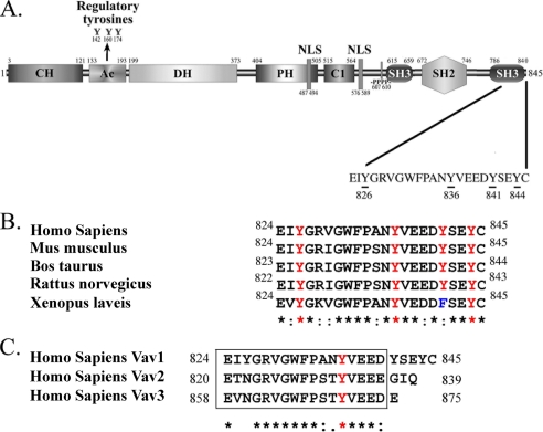
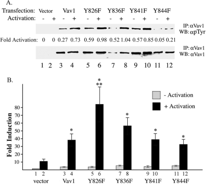
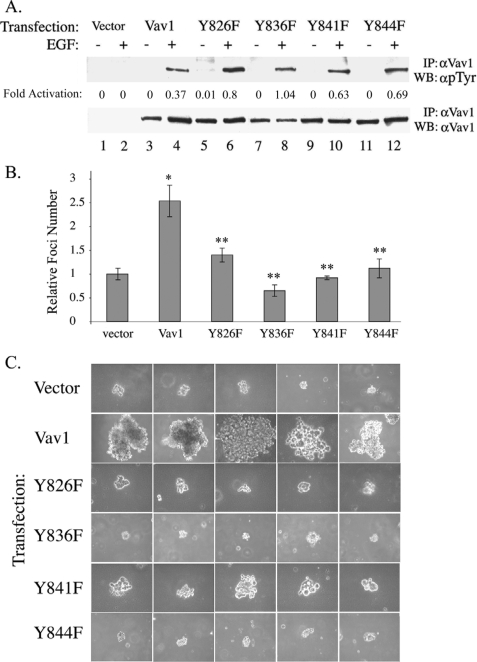
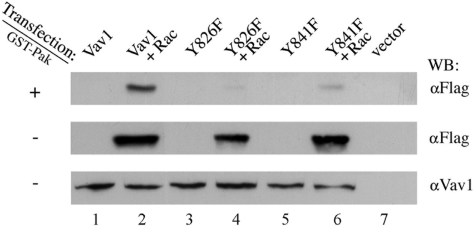
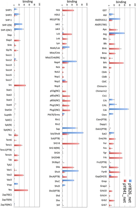
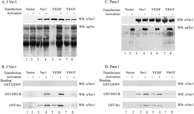
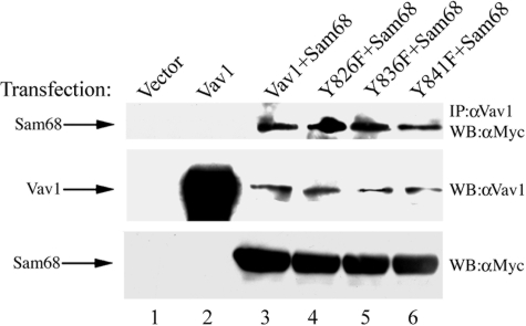
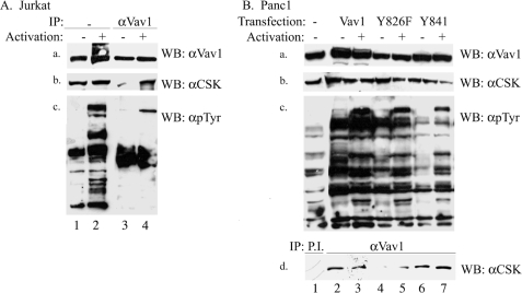
The guanine nucleotide exchange factor (GEF) Vav1 is an essential signal transducer protein in the hematopoietic system, where it is expressed physiologically. It is also involved in several human malignancies. Tyrosine phosphorylation at the Vav1 amino terminus plays a central role in regulating its activity; however, the role of carboxyl terminal tyrosine residues is unknown. We found that mutation of either Tyr-826 (Y826F) or Tyr-841 (Y841F) to phenylalanine led to loss of Vav1 GEF activity. When these Vav1 mutants were ectopically expressed in pancreatic cancer cells lacking Vav1, they failed to induce growth in agar, indicating loss of transforming potential. Furthermore, although Y841F had no effect on Vav1-stimulated nuclear factor of activated T cells (NFAT) activity, Y826F doubled NFAT activity when compared with Vav1, suggesting that Tyr-826 mediates an autoinhibitory effect on NFAT activity. SH2 profiling revealed that Shc, Csk, Abl, and Sap associate with Tyr-826, whereas SH2-B, Src, Brk, GTPase-activating protein, and phospholipase C-gamma associate with Tyr-841. Although the mutations in the Tyr-826 and Tyr-841 did not affect the binding of the carboxyl SH3 of Vav1 to other proteins, binding to several of the proteins identified by the SH2 profiling was lost. Of interest is Csk, which associates with wild-type Vav1 and Y841F, yet it fails to associate with Y826F, suggesting that loss of binding between Y826F and Csk might relieve an autoinhibitory effect, leading to increased NFAT. Our data indicate that GEF activity is critical for the function of Vav1 as a transforming protein but not for NFAT stimulation. The association of Vav1 with other proteins, detected by SH2 profiling, might affect other Vav1-dependent activities, such as NFAT stimulation.


Figures

FIGURE 1.
FIGURE 1.
Molecular structure of Vav1. A, the location of the following Vav1 domains is indicated: calponin-homology (CH) domain; acidic (Ac) motif that contains three tyrosine residues as indicated; a DH domain; a PH domain; a C1 domain; a proline-rich region (–PPPP–); two nuclear localization signals (NLS); and two SH3 domains and an SH2 domain. Tyrosine residues mutated in the current study are underlined, and the number of the residue is indicated underneath. B, sequence alignment of Vav1 among different species. The tyrosine residues mutated in this study are highlighted in human Vav1 in red as well as by a red asterisk. C, sequence alignment of the carboxyl terminus of human Vav1, Vav2, and Vav3.
FIGURE 2.
FIGURE 2.
The effect of mutations in the carboxyl terminus of Vav1 on NFAT stimulation. A, J.Vav1 cells were transiently transfected with vector only (lanes 1 and 2); Vav1 (lanes 3 and 4); Y826F (lanes 5 and 6); Y836F (lanes 7 and 8); Y841F (lanes 9 and 10); or Y844F (lanes 11 and 12). The cells were either nonstimulated (−) or stimulated with anti-CD3 and anti-CD28 (+) as described. Cell lysates were immunoprecipitated (IP) with anti-Vav1 Abs, separated on SDS-PAGE, and immunoblotted (WB) with anti-Tyr(P) (αpTyr) mAbs. After stripping, membranes were reprobed with anti-Vav1 mAbs. Fold activation was determined by dividing the optical density of phosphorylated protein detected in the upper panel by that detected in the lower panel. B, J.Vav1 cells were electroporated with Renilla, NFAT-luciferase reporter gene plasmids, and either empty vector or vector containing Vav1 or Vav1 mutants Y826F, Y836F, Y841F, and Y844F as outlined. Eighteen hours later, cells were activated for 6 h with anti-CD3 and anti-CD28 mAbs and then lysed. Relative luciferase activity indicates the ratio of firefly luc activity to Renilla luc activity. Induction levels are relative to that of the indicated vector alone under control conditions. Histograms represent the mean ± S.E. from five experiments. Statistically significant results when compared with vector alone (lane 2) are indicated by one asterisk, whereas two asterisks indicate significant statistical differences when compared with Vav1 (lane 4).
FIGURE 3.
FIGURE 3.
Vav1 mutants are defective in their ability to enhance Panc1 growth in soft agar. A, Panc1 cells were transiently transfected with vector only (lanes 1 and 2); Vav1 (lanes 3 and 4); Y826F (lanes 5 and 6); Y836F (lanes 7 and 8); Y841F (lanes 9 and 10); or Y844F (lanes 11 and 12). The cells were either nonstimulated (−) or stimulated with EGF (+) as described. Cell lysates were immunoprecipitated (IP) with anti-Vav1 Abs, separated on SDS-PAGE, and immunoblotted (WB) with anti-Tyr(P) (αpTyr) mAbs. After stripping, membranes were reprobed with anti-Vav1 mAbs. Fold activation indicates the division of the phosphorylated protein detected in the upper panel versus its expression detected in the lower panel. B, Panc1 cells were transfected with Vav1 and the various Vav1 mutants, as indicated, according to the protocol detailed under “Experimental Procedures.” Cells were trypsinized 48 h following transfection, suspended in RPMI medium containing 0.3% agar and 10% calf serum, and plated onto a bottom layer containing 0.8% agar. Cells were plated at a density of 1 × 10⁵/well in a 6-well plate, and the number of colonies was counted 14 days later. The number of foci obtained in each experiment was divided by the number of foci in control cells transfected with vector only to obtain relative foci. Histograms show the mean ± S.E. of triplicate values from three independent experiments. Statistically significant results when compared with vector alone are indicated by one asterisk, whereas two asterisks indicate significant statistical differences when compared with Vav1. C, representative photographs of the foci generated in B.
FIGURE 4.
FIGURE 4.
Y826F and Y841F Vav1 mutants lose their capacity to function as GEF for Rac. Panc1 cells were transfected with Vav1 (lane 1), Vav1 and FLAG epitope-tagged Rac (lane 2), Y826F (lane 3), Y826F and FLAG epitope-tagged Rac (lane 4), Y841F (lane 5), Y841F and FLAG epitope-tagged Rac (lane 6), or vector only (lane 7). Cell lysates were then incubated with the bacterial fusion protein that expresses Pak immobilized on glutathione-Sepharose beads. Bound proteins were separated on SDS-PAGE and immunoblotted (WB) with anti-FLAG mAbs (+ panel). Expression of transfected Vav1 (middlepanel) and Rac-FLAG (lowerpanel) were detected by anti-Vav1 and anti-FLAG mAbs as indicated. The figure shows one representative experiment of two experiments performed.
FIGURE 5.
FIGURE 5.
Binding of SH2/PTB domains to potential tyrosine phosphorylation sites of Vav1. A, in vitro SH2/PTB binding assay was performed as described under “Experimental Procedures.” Positions and sequences of Vav1 peptides used in the binding experiments are shown. Peptides were immobilized on a membrane in multiple duplicate arrays as shown in the diagram on the right. pY286, Tyr(P)-286; pY841, Tyr(P)841; B, a representative result of large scale screen is shown. There are three types of control probes: avidin as a spotting control for biotinylated peptide; and anti-Tyr(P) (anti-pTyr) as a positive control for tyrosine phosphorylation; GST probes as negative controls. SH2 domains of ShcA, ShcC, Csk, and Sap families bound strongly to Tyr(P)-826, whereas SH2 domains of SH2-B and Src families bound to Tyr(P)-841. C, summary of quantitated screening data. Mean binding intensities ± S.E. for each SH2 domain family to respective peptides are shown. See supplemental material for more details.
FIGURE 6.
FIGURE 6.
Quantified SH2/PTB screening data. The signal intensities of multiple independent experiments with duplicate spots were quantified using densitometry, and the net binding intensity was obtained by subtracting the value of binding to unphosphorylated peptide from the value of binding to phosphorylated peptide. The bar graph indicates the mean value and S.E. for each SH2/PTB domain probe.
FIGURE 7.
FIGURE 7.
Binding of Vav1, Y826F, and Y841F to SH2-B and Src. A, J.Vav1 cells were transiently transfected with vector only (lanes 1 and 2); Vav1 (lanes 3 and 4); Y826F (lanes 5 and 6); or Y841F (lanes 7 and 8). The cells were either nonstimulated (−; lanes 1, 3, 5, and 7) or stimulated with anti-CD3 and anti-CD28 mAbs as described (+; lanes 2, 4, 6, and 8). Cell lysates were separated on SDS-PAGE and immunoblotted (WB) with anti-Vav1 mAbs (upper panel), and after stripping of the membrane, with Tyr(P) (αpTyr) mAbs (lower panel). B, lysates of J.Vav1 cells described in A were incubated with bacterial fusion proteins (GST-G830V, GST-SH2-B, or GST-Src) and then immobilized on glutathione-Sepharose beads. Bound proteins were resolved on SDS-PAGE gels and immunoblotted with anti-Vav1 mAbs. C, Panc1 cells were transiently transfected with vector only (lanes 1 and 2); Vav1 (lanes 3 and 4); Y826F (lanes 5 and 6); or Y841F (lanes 7 and 8). The cells were either nonstimulated (−; lanes 1, 3, 5, and 7) or stimulated with EGF as described (+; lanes 2, 4, 6, and 8). Cell lysates were separated on SDS-PAGE and immunoblotted with anti-Vav1 mAbs (upper panel), and after stripping the membrane, with Tyr(P) mAbs (lower panel). D, lysates of J.Vav1 cells described in A were incubated with bacterial fusion proteins (GST-G830V, GST-SH2-B, or GST-Src) and then immobilized on glutathione-Sepharose beads. Bound proteins were resolved on SDS-PAGE gels and immunoblotted with anti-Vav1 mAbs.
FIGURE 8.
FIGURE 8.
Vav1 mutants interact with Sam68. HEK293T cells were transfected with the following plasmids: an empty vector; Vav1; and Vav1, Y826F, Y836F, and Y841F together with Myc-tagged Sam68, as indicated. The transfected cells were lysed, immunoprecipitated (IP) with anti-Vav1 Abs, and immunoblotted (WB) with anti-Myc mAbs (upper panel). The level of Vav1 or Sam68 in the cell lysates was determined by immunoblotting with either anti-Vav1 mAbs (middle panel) or anti-Myc mAbs (lower panel).
FIGURE 9.
FIGURE 9.
Binding of Vav1, Y826F, and Y841F to CSK in vivo. A, Jurkat T cells were activated with anti-CD3 Abs and anti-CD28 mAbs as described under “Experimental Procedures” (+, lanes 2 and 4) or left nonactivated (−, lanes 1 and 3) and then lysed and immunoprecipitated (IP) with anti-Vav1 Abs (lanes 3 and 4). or nonimmunoprecipitated (lanes 1 and 2). Immunoblotting (WB) was performed with anti-Vav1 Abs (panel a), anti-CSK Abs (panel b), or anti-Tyr(P) Abs (panel c). B, Panc1 cells were transiently transfected with wild-type Vav1 only (lanes 1 and 2); Y826F (lanes 3 and 4); or Y841F (lanes 5 and 6). The cells were either nonstimulated (−; lanes 1, 2, 4, and 6) or stimulated with EGF as described (+; lanes 3, 5, and 7). Cell lysates were separated on SDS-PAGE and immunoblotted with anti-Vav1 mAbs (panel a), anti-CSK Abs (panel b), and anti-Tyr(P) Abs (panel c). Lysates of these cells were immunoprecipitated with preimmune sera (P.I.; lane 1; panel d) or anti-Vav1 Abs (lanes 2–7; panel d). Immunoblotting was performed with anti-CSK Abs. To avoid unmasking of CSK, in A (panel b) and B (panel d), we used monoclonal anti-rabbit IgG and native peroxidase antibody produced in mouse (clone RabT-50, Sigma).

Similar articles

Cited by

References

    1. Sjöblom T., Jones S., Wood L. D., Parsons D. W., Lin J., Barber T. D., Mandelker D., Leary R. J., Ptak J., Silliman N., Szabo S., Buckhaults P., Farrell C., Meeh P., Markowitz S. D., Willis J., Dawson D., Willson J. K., Gazdar A. F., Hartigan J., Wu L., Liu C., Parmigiani G., Park B. H., Bachman K. E., Papadopoulos N., Vogelstein B., Kinzler K. W., Velculescu V. E. (2006) Science 314, 268–274 - PubMed
    1. Hunter T. (2000) Cell 100, 113–127 - PubMed
    1. Schlessinger J. (2000) Cell 103, 211–225 - PubMed
    1. Manning G., Whyte D. B., Martinez R., Hunter T., Sudarsanam S. (2002) Science 298, 1912–1934 - PubMed
    1. Alonso A., Sasin J., Bottini N., Friedberg I., Friedberg I., Osterman A., Godzik A., Hunter T., Dixon J., Mustelin T. (2004) Cell 117, 699–711 - PubMed

Publication types

MeSH terms