. 2010 Jul 1;588(Pt 13):2403-15.
doi: 10.1113/jphysiol.2010.187096. Epub 2010 May 10.

Electrical and chemical synapses between relay neurons in developing thalamus

Affiliations

Electrical and chemical synapses between relay neurons in developing thalamus

Seung-Chan Lee et al. J Physiol. .

Abstract

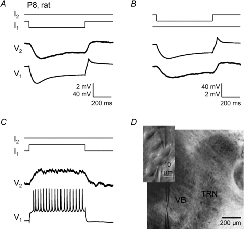
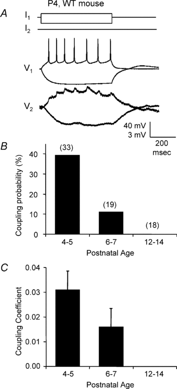
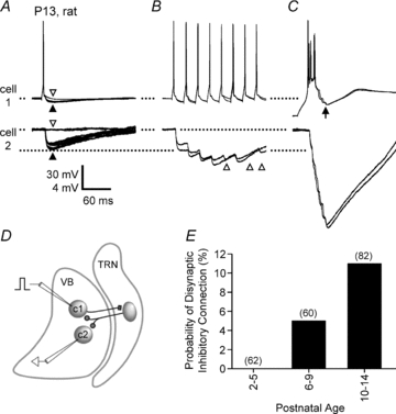
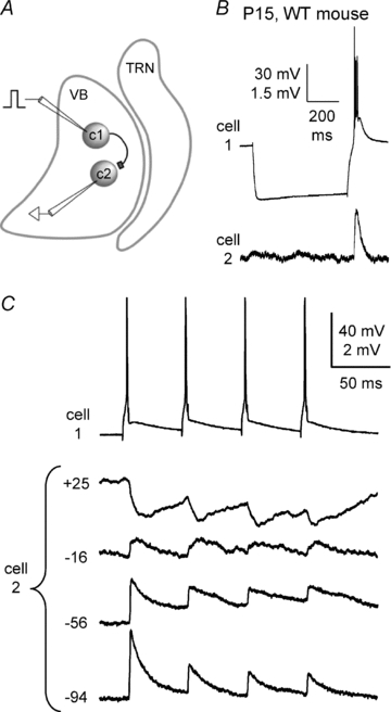
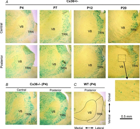
Gap junction-mediated electrical synapses interconnect diverse types of neurons in the mammalian brain, and they may play important roles in the synchronization and development of neural circuits. Thalamic relay neurons are the major source of input to neocortex. Electrical synapses have not been directly observed between relay neurons in either developing or adult animals. We tested for electrical synapses by recording from pairs of relay neurons in acute slices of developing ventrobasal nucleus (VBN) of the thalamus from rats and mice. Electrical synapses were common between VBN relay neurons during the first postnatal week, and then declined sharply during the second week. Electrical coupling was reduced among cells of connexin36 (Cx36) knockout mice; however, some neuron pairs remained coupled. This implies that electrical synapses between the majority of coupled VBN neurons require Cx36 but that other gap junction proteins also contribute. The anatomical distribution of a beta-galactosidase reporter indicated that Cx36 was expressed in some VBN neurons during the first postnatal week and sharply declined over the second week, consistent with our physiological results. VBN relay neurons also communicated via chemical synapses. Rare pairs of relay neurons excited one another monosynaptically. Much more commonly, spikes in one relay neuron evoked disynaptic inhibition (via the thalamic reticular nucleus) in the same or a neighbouring relay neuron. Disynaptic inhibition between VBN cells emerged as electrical coupling was decreasing, during the second postnatal week. Our results demonstrate that thalamic relay neurons communicate primarily via electrical synapses during early postnatal development, and then lose their electrical coupling as a chemical synapse-mediated inhibitory circuit matures.


Figures

Figure 1
Figure 1. Electrical synapses between rat VBN neurons
Electrical coupling recorded in current clamp from a pair of closely spaced VBN neurons (P8 rat). A, negative intracellular current steps (−150 pA) in cell 1 evoked a large voltage hyperpolarzation in cell 1 and a smaller hyperpolarization, due to coupling, in cell 2 (coupling coefficient ∼0.04). B, the same cell pair, but in this case negative current steps (−100 pA) were delivered to cell 2. C, positive current steps (+340 pA) delivered to cell 1 produced depolarization and spiking in cell 1 and smaller depolarization in cell 2. Calibration bars in A also apply to C. D, IR-DIC image of the paired whole-cell recording in the ventrobasal nucleus (VBN) of a thalamocortical slice. Inset shows a higher power view of the recorded cells.
Figure 3
Figure 3. Electrical synapses between VBN neurons of WT mice
A, recording of an electrically coupled VBN neuronal pair from a P4 wild-type mouse. Current pulses of 25 pA and −50 pA were injected into the first cell. Note the postsynaptic ‘spikelets’ in the strongly coupled paired cell (CC = 0.079). B and C, coupling probabilities and coefficients of VBN neurons in wild-type mice at different postnatal ages. Values in parentheses in B are the number of pairs recorded for the respective age groups.
Figure 2
Figure 2. Developmental change in electrical coupling between rat VBN neurons
A, the probability of observing electrical coupling between adjacent VBN cells decreased with postnatal age (P2–P31). Values in parentheses are the number of pairs recorded for the respective age groups. B, coupling coefficients of electrically coupled pairs also decreased with postnatal age (P < 0.02, linear regression). Circles represent individual pairs and dashes represent group means. C, estimated junctional conductances of connected pairs from P2 to P14 (P= 0.051, linear regression).
Figure 7
Figure 7. Disynaptic inhibitory transmission between VBN neurons
A–D, disynaptic inhibition in a pair of VBN cells from a P13 rat. Spikes were evoked in cell 1 (with intracellular current steps) while disynaptic IPSPs were recorded in cell 2 and in cell 1. Cell 2 was maintained at a resting potential of −66 mV. A, single spikes in cell 1 evoked 3–4 mV IPSPs in cell 2 (filled arrowhead), although failures occurred occasionally (2 failures in 15 trials shown; open arrowhead). Spikes in cell 1 also evoked IPSPs in cell 1 (filled arrowhead); these are evident by comparison with trials where the IPSP failed (open arrowhead). The IPSP failures in cell 1 occurred on the same trials as those in cell 2. B, trains of spikes at 40 Hz resulted in IPSP summation. Two example trains are shown. IPSPs were evoked in cell 2 following most spikes in cell 1 (IPSP failures are evident after spikes 5, 7 and 8: open arrowheads). The individual IPSPs depressed in amplitude during the train, but summation caused peak voltages to be nearly 2-fold larger than single spike-evoked IPSPs. C, bursts were most effective at evoking disynaptic inhibition. Two bursts are shown (4 spikes per burst). Cell 1 was first stepped to −90 mV with hyperpolarizing current (>500 ms), then current was removed, allowing for depolarization and bursting. Peak burst-evoked IPSPs recorded in cell 2 were larger than peak train-evoked IPSPs in B. Note the presence of IPSPs also in cell 1 following the burst (arrow). D, schematic of the presumed connections underlying the example of A–C. Cell 1 (c1) in VBN makes excitatory synapses with at least one inhibitory cell in TRN (not recorded). The TRN cell makes inhibitory synapses with cells 1 and 2 in the VBN, leading to IPSPs. E, the probability of observing disynaptic inhibition between adjacent VBN cells across early postnatal development in rats. Values in parentheses are the number of connections tested for the respective age groups (2 connections tested per pair).
Figure 6
Figure 6. Excitatory chemical synaptic transmission between VBN neurons
Recordings from a pair of VBN cells from a P15 mouse. A, schematic diagram of protocol and presumed connections for the example in B and C. Cell 1 makes an excitatory synapse with cell 2. Intracellular current steps were injected into cell 1 (c1) evoking spikes. Cell 1 projected to cell 2 (c2), producing monosynaptic EPSPs. B, initially, long negative current pulses (600 ms) were injected into cell 1, producing hyperpolarization followed by offset bursts. The spike bursts in cell 1 evoked EPSPs in cell 2. This cell pair was not electrically coupled. C, reversal potential of the EPSPs was tested by inducing spike trains in cell 1 (4 spikes, 20 Hz) and recording EPSPs in cell 2 while its steady-state membrane potential was adjusted across a wide range (−94 to +25 mV) with intracellular current. The EPSPs reversed near 0 mV, consistent with glutamatergic synapses.
Figure 4
Figure 4. Most electrical coupling between young mouse VBN neurons depends on Cx36
A, coupling probabilities in VBN nuclei of KO vs. WT mice at P4–5. Coupling probability was reduced in Cx36 KO. Values in parentheses are the number of pairs recorded for each genotype. B, coupling coefficients of VBN cell pairs from WT and Cx36 KO mice. Shading demarcates coupling coefficients below threshold (CC < 0.01, see Methods). C, example traces from one of the 4 electrically coupled VBN pairs from the Cx36 KO group (P4, CC = 0.023). Negative current pulses (−80 pA) were injected into cell 1, causing a large hyperpolarization in that cell and a smaller hyperpolarization, due to coupling, in cell 2.
Figure 5
Figure 5. β-Galactosidase reporter expression for Cx36 in mouse VBN at different postnatal ages
A, images of β-galactosidase staining in thalamic slices from Cx36 heterozygote mice (Cx36+/−) at different postnatal ages. Bottom row images are from the posterior part of VBN, and top row are from the central part of VBN (i.e. more anterior). The inset in the lower-right region shows the boxed VBN region of the posterior P20 section at higher magnification. B, β-galactosidase signal in thalamic slices from a P4 Cx36 KO mouse (homozygote, Cx36−/−). C, β-galactosidase signal in a thalamic slice from a P4 WT mouse (Cx36+/+). Dotted lines outline approximate boundaries of VBN and TRN.

Similar articles

Cited by

References

    1. Agmon A, Connors BW. Thalamocortical responses of mouse somatosensory (barrel) cortex in vitro. Neuroscience. 1991;41:365–379. - PubMed
    1. Amitai Y, Gibson JR, Beierlein M, Patrick SL, Ho AM, Connors BW, Golomb D. The spatial dimensions of electrically coupled networks of interneurons in the neocortex. J Neurosci. 2002;22:4142–4152. - PMC - PubMed
    1. Arumugam H, Liu X, Colombo PJ, Corriveau RA, Belousov AB. NMDA receptors regulate developmental gap junction uncoupling via CREB signalling. Nat Neurosci. 2005;8:1720–1726. - PubMed
    1. Belluardo N, Mudo G, Trovato-Salinaro A, Le Gurun S, Charollais A, Serre-Beinier V, Amato G, Haefliger JA, Meda P, Condorelli DF. Expression of connexin36 in the adult and developing rat brain. Brain Res. 2000;865:121–138. - PubMed
    1. Bennett MV. Physiology of electrotonic junctions. Ann N Y Acad Sci. 1966;137:509–539. - PubMed

Publication types

MeSH terms

LinkOut - more resources