. 2010 Jun 7;207(6):1135-43.
doi: 10.1084/jem.20092301. Epub 2010 May 10.

Complement drives Th17 cell differentiation and triggers autoimmune arthritis

Affiliations

Complement drives Th17 cell differentiation and triggers autoimmune arthritis

Motomu Hashimoto et al. J Exp Med. .

Abstract

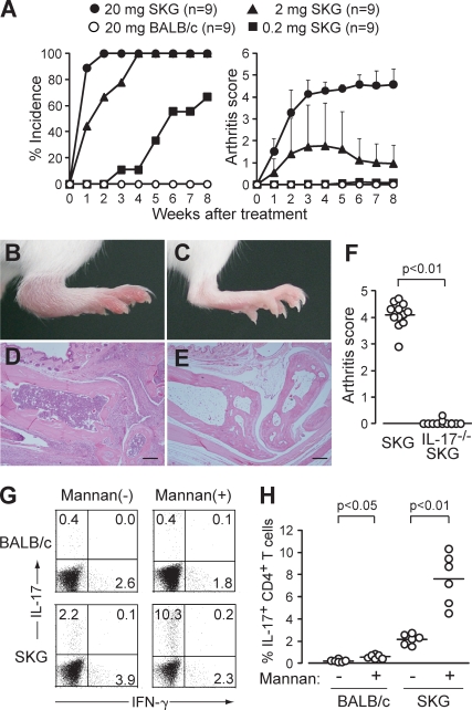
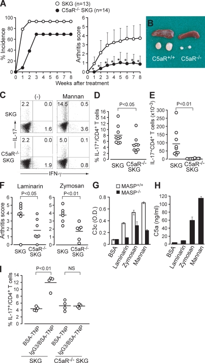
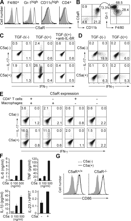
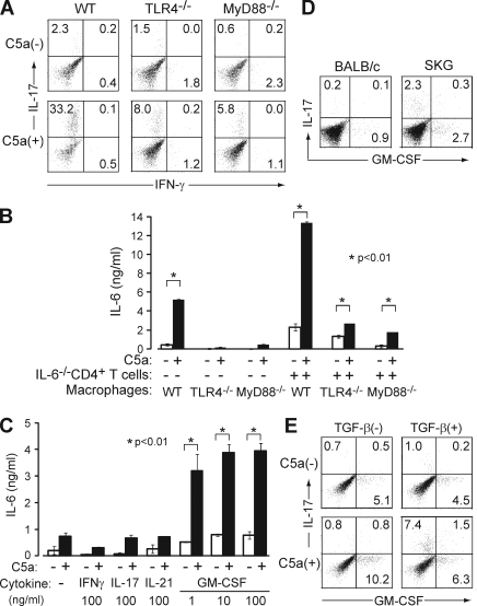
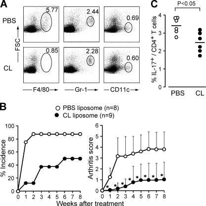
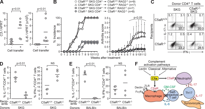
Activation of serum complement triggers Th17 cell-dependent spontaneous autoimmune disease in an animal model. In genetically autoimmune-prone SKG mice, administration of mannan or beta-glucan, both of which activate serum complement, evoked Th17 cell-mediated chronic autoimmune arthritis. C5a, a chief component of complement activation produced via all three complement pathways (i.e., lectin, classical, and alternative), stimulated tissue-resident macrophages, but not dendritic cells, to produce inflammatory cytokines including IL-6, in synergy with Toll-like receptor signaling or, notably, granulocyte/macrophage colony-stimulating factor (GM-CSF). GM-CSF secreted by activated T cells indeed enhanced in vitro IL-6 production by C5a-stimulated macrophages. In vivo, C5a receptor (C5aR) deficiency in SKG mice inhibited the differentiation/expansion of Th17 cells after mannan or beta-glucan treatment, and consequently suppressed the development of arthritis. Transfer of SKG T cells induced Th17 cell differentiation/expansion and produced arthritis in C5aR-sufficient recombination activating gene (RAG)-/- mice but not in C5aR-deficient RAG-/- recipients. In vivo macrophage depletion also inhibited disease development in SKG mice. Collectively, the data suggest that complement activation by exogenous or endogenous stimulation can initiate Th17 cell differentiation and expansion in certain autoimmune diseases and presumably in microbial infections. Blockade of C5aR may thus be beneficial for controlling Th17-mediated inflammation and autoimmune disease.


Figures

Figure 1.
Figure 1.
Mannan triggers autoimmune arthritis by expanding Th17 cells. (A) Joint score of 8∼12-wk-old SKG or BALB/c mice that received a single i.p. injection of mannan at the indicated doses. A total of two independent experiments are shown. Error bars are means ± SD of scores. (B–E) A representative joint swelling and histology of an SKG (B and D) and a BALB/c mouse (C and E) 8 wk after mannan treatment (hematoxylin and eosin staining). Bars, 200 µm. (F) Joint scores of IL-17+/+ or IL-17−/− SKG mice 8 wk after mannan treatment (n = 12). Horizontal bars are the means of each group. (G) Intracellular staining of IL-17 and IFN-γ in CD4+ T cells in the popliteal lymph nodes from SKG or BALB/c mice 8 wk after mannan treatment (numbers indicate percentages). One representative staining out of six independent experiments is shown. (H) Percentages of IL-17+ cells in CD4+ T cells in each SKG or BALB/c group (n = 6), as shown in G. Horizontal bars are the means of each group.
Figure 2.
Figure 2.
C5a drives Th17 cell differentiation and triggers arthritis. (A) Joint score of C5aR+/+ or C5aR−/− SKG mice treated by 20 mg mannan. A total of two independent experiments are shown. Error bars are means ± SD of scores. *, P < 0.05. (B) Spleen and popliteal lymph nodes of C5aR+/+ or C5aR−/− SKG mice 8 wk after mannan treatment. (C) Intracellular staining of IL-17 and IFN-γ in CD4+ T cells in the popliteal lymph nodes of C5aR+/+ or C5aR −/− SKG mice, as shown in B (numbers indicate percentages). One representative staining out of nine independent experiments is shown. (D and E) Percentages (D) and absolute numbers (E) of IL-17+ cells in CD4+ T cells in each group (n = 9), as shown in C. (F) Joint scores of C5aR+/+ or C5aR−/− SKG mice 8 wk after treatment by laminarin (n = 7) or zymosan (n = 6). (G) C3 deposition assay with laminarin-, zymosan-, or mannan-coated wells incubated with 2% sera from MASP-intact or -null C57BL/6 sera. (H) C5a ELISA for the supernatant of the C3 deposition assay with BALB/c sera, as shown in G. Error bars are means ± SD. (I) C5aR+/+ and C5aR−/− SKG mice were i.p. injected with TNP-BSA alone or IgG3 anti-TNP/TNP-BSA IC. The percentage of IL-17+ cells in the peritoneal CD4+ T cells was assessed on day 7 (n = 4). Horizontal bars are the means.
Figure 3.
Figure 3.
C5a acts on tissue-resident macrophages to facilitate Th17 cell differentiation. (A) F4/80+ cells in the peritoneal cavity, and Gr-1high, CD11chigh, and CD4+ cells in the spleen of nontreated SKG mice were stained for C5aR (continuous line) or isotype control (shading). (B) Cells infiltrating in the arthritic joint of mannan-treated SKG mice were stained for C5aR, CD11b, Gr-1, and F4/80. C5aR+ CD11b+ cells are shown for the expression of Gr-1 and F4/80. (C) BALB/c CD4+ T cells were stimulated with anti-CD3 and cultured with resident peritoneal macrophages in the presence or absence of TGF-β, recombinant C5a, or anti–IL-6R, and stained for intracellular cytokines on day 3. (D) Intracellular cytokine staining of BALB/c CD4+ T cells cultured with splenic DCs in the presence or absence of TGF-β and C5a. (E) Intracellular cytokine staining of CD4+ T cells after criss-cross co-culture with C5aR+/+ or C5aR−/− CD4+ T cells and C5aR+/+ or C5aR−/− macrophages in the presence of TGF-β. (F) ELISA assessment (triplicates) for IL-6, TNF, and IL-1β produced by macrophages, or quantitative RT-PCR for their IL-23 mRNA expression, when macrophages were stimulated overnight by C5a at the indicated doses. Error bars are means ± SD. (G) CD86 expression on C5aR+/+ or C5aR−/− macrophages cultured with or without C5a for 4 h. Shading indicates isotype control. Results in A–G represent three independent experiments. Numbers in B–E indicate percentages.
Figure 4.
Figure 4.
C5a drives Th17 cell differentiation in synergy with TLR or GM-CSF. (A) C57BL/6 CD4+ T cells were cultured with TLR4−/−, MyD88−/−, or wild-type C57BL/6 macrophages in the presence of TGF-β with or without C5a. (B) Macrophages alone from these mice or co-cultured with anti-CD3–stimulated CD4+ T cells from IL-6−/− mice were stimulated with C5a overnight. IL-6 in the supernatant was determined by ELISA. (C) MyD88−/− macrophages were stimulated by C5a overnight in the presence or absence of cytokines at the indicated doses. IL-6 in the supernatant was determined by ELISA (triplicates). Error bars are means ± SD. (D) Freshly isolated BABL/c or SKG splenic CD4+ T cells were stained for intracellular IL-17 and GM-CSF. (E) BALB/c CD4+ T cells were cultured with macrophages in the presence or absence of C5a and/or TGF-β, stimulated with anti-CD3, and stained for intracellular cytokines on day 3. Results in A–E represent three independent experiments. Numbers in A, D, and E indicate percentages.
Figure 5.
Figure 5.
Macrophages are required for in vivo Th17 expansion and induction of arthritis. (A) Specific depletion of macrophages in SKG mice 3 d after i.v. injection of 200 µl CL. One representative out of three independent experiments is shown (numbers indicate percentages). (B) Joint scores of SKG mice i.v. injected with 200 µl PBS or CL 1 d before mannan treatment. *, P < 0.05. Error bars are means ± SD. (C) Percentage of IL-17+ CD4+ T cells in the popliteal lymph nodes of SKG mice 2 wk after mannan treatment. Mice were pretreated by an i.v. injection of PBS or CL (n = 6). Horizontal bars are the means.
Figure 6.
Figure 6.
C5a promotes spontaneous differentiation of CD4+ T cells to Th17 cells via homeostatic proliferation. (A) Quantitative RT-PCR for C3 and IL-6 mRNA in the joints of RAG2−/− mice 12 wk after transfer of SKG CD4+ T cells (n = 6). (B) 106 C5aR+/+ or C5aR−/− SKG CD4+ T cells were transferred to C5aR+/+ or C5aR−/− RAG2−/− mice. Joint scores were assessed every week in two independent experiments. Error bars are means ± SD. (C) 2 × 10⁶ C5aR+/+ or C5aR−/− SKG CD4+ T cells were transferred to C5aR+/+ or C5aR−/− RAG2−/− mice. Intracellular cytokines in recipient splenic CD4+ T cells were stained on day 7 (numbers indicate percentages). One representative out of four independent experiments is shown. (D and E) Percentages of IL-17+ or IFN-γ+ cells among CD4+ T cells in C5aR+/+ or C5aR−/− RAG2−/− mice (n = 4 each) after transfer of SKG (D) or BALB/c CD4+ T cells (E). (F) A model for the role of complement activation in Th17-mediated autoimmune arthritis. Horizontal bars in A, D, and E are the means.

References

    1. Díaz de Ståhl T., Dahlstrom J., Carroll M.C., Heyman B. 2003. A role for complement in feedback enhancement of antibody responses by IgG3. J. Exp. Med. 197:1183–1190 10.1084/jem.20022232 - DOI - PMC - PubMed
    1. Fang C., Zhang X., Miwa T., Song W.C. 2009. Complement promotes the development of inflammatory T-helper 17 cells through synergistic interaction with Toll-like receptor signaling and interleukin-6 production. Blood. 114:1005–1015 10.1182/blood-2009-01-198283 - DOI - PMC - PubMed
    1. Fujita T. 2002. Evolution of the lectin-complement pathway and its role in innate immunity. Nat. Rev. Immunol. 2:346–353 10.1038/nri800 - DOI - PubMed
    1. Garlatti V., Belloy N., Martin L., Lacroix M., Matsushita M., Endo Y., Fujita T., Fontecilla-Camps J.C., Arlaud G.J., Thielens N.M., Gaboriaud C. 2007. Structural insights into the innate immune recognition specificities of L- and H-ficolins. EMBO J. 26:623–633 10.1038/sj.emboj.7601500 - DOI - PMC - PubMed
    1. Grabstein K.H., Urdal D.L., Tushinski R.J., Mochizuki D.Y., Price V.L., Cantrell M.A., Gillis S., Conlon P.J. 1986. Induction of macrophage tumoricidal activity by granulocyte-macrophage colony-stimulating factor. Science. 232:506–508 10.1126/science.3083507 - DOI - PubMed

Publication types

MeSH terms