. 2010 Jun 7;207(6):1223-34.
doi: 10.1084/jem.20092170. Epub 2010 May 10.

Peptide-MHC heterodimers show that thymic positive selection requires a more restricted set of self-peptides than negative selection

Affiliations

Peptide-MHC heterodimers show that thymic positive selection requires a more restricted set of self-peptides than negative selection

Jeremy Juang et al. J Exp Med. .

Abstract

T cell selection and maturation in the thymus depends on the interactions between T cell receptors (TCRs) and different self-peptide-major histocompatibility complex (pMHC) molecules. We show that the affinity of the OT-I TCR for its endogenous positively selecting ligands, Catnb-H-2Kb and Cappa1-H-2Kb, is significantly lower than for previously reported positively selecting altered peptide ligands. To understand how these extremely weak endogenous ligands produce signals in maturing thymocytes, we generated soluble monomeric and dimeric peptide-H-2Kb ligands. Soluble monomeric ovalbumin (OVA)-Kb molecules elicited no detectable signaling in OT-I thymocytes, whereas heterodimers of OVA-Kb paired with positively selecting or nonselecting endogenous peptides, but not an engineered null peptide, induced deletion. In contrast, dimer-induced positive selection was much more sensitive to the identity of the partner peptide. Catnb-Kb-Catnb-Kb homodimers, but not heterodimers of Catnb-Kb paired with a nonselecting peptide-Kb, induced positive selection, even though both ligands bind the OT-I TCR with detectable affinity. Thus, both positive and negative selection can be driven by dimeric but not monomeric ligands. In addition, positive selection has much more stringent requirements for the partner self-pMHC.


Figures

Figure 1.
Figure 1.
Affinity of naturally occurring positively selecting peptide-Kb ligands for the OT-I TCR. Equilibrium binding of soluble monomeric OVA-Kb, Catnb-Kb, and Cappa1-Kb to OT-I TCR measured by Biacore 3000 SPR at 10°C at various concentrations of peptide-Kb. Error bars show SE from three to five separate runs. The table indicates the name, the sequence of the peptide, the affinity (KD) in micromolar, the SE (± SE), and the goodness of fit (R2). The results are representative of at least three independent experiments. RU, relative units.
Figure 2.
Figure 2.
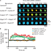
Calcium signaling in thymocytes in response to high-affinity pMHC. Soluble homodimers of agonist pMHCs can stimulate calcium signaling in thymocytes, whereas monomers of agonist pMHC cannot. (A) OT-I TCR transgenic Kb−/−Db−/− thymocytes were loaded with fura-2, a ratiometric calcium-sensitive fluorescent dye (Tsien, 1989), and mixed with soluble OVA-Kb monomers (1 mg/ml or 17 µM) or OVA-Kb—OVA-Kb homodimers (100 µg/ml or 0.87 µM). Representative Ca2+ ratio images obtained with fura-2 (340/380 nm) are shown in false color scale as indicated in the bottom right corner. (B) Fura-2 ratios (340/380 nm) were measured every 15 s for thymocytes in randomly chosen microscope fields, and their mean Ca2+ ratio was plotted as a function of time (fura ratio mean ± SEM; n = 7). The results are representative of at least three independent experiments.
Figure 3.
Figure 3.
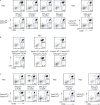
Negative selection in FTOCs in response to dimeric pMHC. Agonist monomers cannot elicit negative selection in thymocytes, but agonist homodimers and agonist-self-peptide heterodimers induce negative selection in FTOC at a similar concentration. (A) Thymic lobes were excised from OT-I Kb−/−Db−/− embryos at gestational day 16. One lobe from each pair was cultured with PBS, whereas the other was cultured with monomers of OVA-Kb (with the cross-linker attached) at 50 µg/ml (0.43 µM). Lobes were harvested after 4 d of culture, stained with anti-CD4 and anti-CD8β, and analyzed by flow cytometry. Representative FACS plots are shown from three or more experiments per condition. (B) Thymic lobes were excised from OT-I Kb−/−Db−/− embryos at gestational day 16 and co-cultured with homodimers of OVA-Kb–OVA-Kb, or with heterodimers OVA-Kb–Catnb-Kb, OVA-Kb–Cappa1-Kb, OVA-Kb–Mapk1-Kb, and OVA-Kb–Stat3-Kb at 5 µg/ml (0.043 µM). Lobes were harvested after 4 d of culture, stained with anti-CD4 and anti-CD8β antibodies, and evaluated by flow cytometry. Representative FACS plots are shown from three or more experiments per condition. (C) Quantification of the data at different concentrations. The percent decrease in the number of CD4+CD8+ DP thymocytes was determined by dividing the difference in the percent of CD4+CD8+ DP population of the experimental lobe by the percentage of CD4+CD8+ DPs in the control lobe and multiplying by 100. Data are presented as mean ± SEM of three or more independent experiments.
Figure 4.
Figure 4.
Calcium signaling in thymocytes in response to low-affinity dimeric pMHC. Soluble homodimers of Catnb-Kb and Cappa1-Kb can stimulate calcium signaling in thymocytes, whereas Catnb-Kb monomers and Mapk1-Kb homodimers and Stat3-Kb homodimers cannot. (A) OT-I TCR transgenic Kb−/−Db−/− thymocytes were loaded with fura-2 and mixed with soluble Catnb-Kb monomers at 1 mg/ml (17 µM) or with Catnb-Kb–Catnb-Kb, Cappa1-Kb–Cappa1-Kb, Mapk1-Kb–Mapk1-Kb, or Stat3-Kb–Stat3-Kb homodimers at 1 mg/ml (8.7 µM). Representative Ca2+ ratio images were obtained with fura-2 (340/380 nm) and are shown using a false color scale as indicated in the bottom right corner. (B) Fura-2 ratios (340/380 nm) were measured every 15 s for thymocytes in randomly chosen microscopic fields and the mean Ca2+ ratio was plotted as a function of time (fura ratio mean ± SEM; n = 7). The results are representative of at least three independent experiments.
Figure 5.
Figure 5.
Positive selection in FTOCs in response to low-affinity dimeric pMHC. Soluble dimers of particular combinations of self-peptide–Kb molecules induce positive selection in OT-I Kb−/−Db−/− FTOC. Thymic lobes were excised from OT-I Kb−/−Db−/− embryos at gestational day 16 and co-cultured with monomers and dimers of endogenous pMHCs at the indicated concentrations. Lobes were harvested after 4 d of culture, stained with anti-CD4 and anti-CD8β antibodies, and evaluated by flow cytometry. (A) These representative FACS plots compare lobes treated with monomeric Catnb-Kb (with the cross-linker attached) to lobes treated with dimeric Catnb-Kb–Catnb-Kb at the indicated protein concentrations. (B) Representative flow cytometry plots comparing lobes treated with a high concentration (1,200 µg/ml or 10 µM) of homodimers (Stat3-Kb–Stat3-Kb, Mapk1-Kb–Mapk1-Kb, or Cappa1-Kb–Cappa1-Kb). (C) Representative flow cytometry plots comparing lobes treated with heterodimers (Catnb-Kb–Cappa1-Kb, Catnb-Kb–Mapk1-Kb, or Catnb-Kb–Stat3-Kb) at the indicated protein concentrations. The results are representative of at least three independent experiments.
Figure 6.
Figure 6.
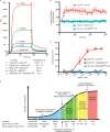
TCR affinity determines the thymic selection outcome, even for very weak ligands. (A) Sensorgram of the interaction between immobilized OT-I TCR at high density with 5 µM Catnb-Kb–Catnb-Kb, Cappa1-Kb–Cappa1-Kb, Mapk1-Kb–Mapk1-Kb, Stat3-Kb–Stat3-Kb, and SIAAFASL-Kb–SIAAFASL-Kb homodimeric pMHC. (B) Mean Ca2+ ratio of thymocytes mixed with 100 µg/ml (0.87 µM) of OVA-Kb–OVA-Kb, OVA-Kb–SIAAFASL-Kb, or OVA-Kb–SIINFEK(BIO)L-Kb. (C) Thymic lobes were excised from OT-I Kb−/−Db−/− embryos at gestational day 16 and co-cultured with homodimers of OVA-Kb–OVA-Kb or with heterodimers OVA-Kb–SIAAFASL-Kb and OVA-Kb–SIINFEK(BIO)L-Kb at various concentrations. Lobes were harvested after 4 d of culture, stained with anti-CD4 and anti-CD8β antibodies, and evaluated by flow cytometry. The percent decrease in the number of CD4+ CD8+ DP thymocytes was determined by dividing the difference in the percentage of CD4+CD8+ DP population of the experimental lobe by the percent of CD4+CD8+ DPs in the control lobe and multiplying by 100. Data are presented as mean ± SEM of three or more independent experiments. (D) A graph summarizing the relationship between pMHC-TCR affinity and thymic selection outcomes. Steady-state KDs measured at 10°C of monomeric and dimeric peptide-Kb binding to OT-I TCR are indicated in the bottom of the graph. * and ‡ represent monomeric KD measurements at 25°C that were obtained previously by Alam et al. (1996) and Alam et al. (1999), respectively.

References

    1. Alam S.M., Travers P.J., Wung J.L., Nasholds W., Redpath S., Jameson S.C., Gascoigne N.R. 1996. T-cell-receptor affinity and thymocyte positive selection. Nature. 381:616–620 10.1038/381616a0 - DOI - PubMed
    1. Alam S.M., Davies G.M., Lin C.M., Zal T., Nasholds W., Jameson S.C., Hogquist K.A., Gascoigne N.R., Travers P.J. 1999. Qualitative and quantitative differences in T cell receptor binding of agonist and antagonist ligands. Immunity. 10:227–237 10.1016/S1074-7613(00)80023-0 - DOI - PubMed
    1. Altman J.D., Reay P.A., Davis M.M. 1993. Formation of functional peptide complexes of class II major histocompatibility complex proteins from subunits produced in Escherichia coli. Proc. Natl. Acad. Sci. USA. 90:10330–10334 10.1073/pnas.90.21.10330 - DOI - PMC - PubMed
    1. Baldwin K.K., Reay P.A., Wu L., Farr A., Davis M.M. 1999. A T cell receptor–specific blockade of positive selection. J. Exp. Med. 189:13–24 10.1084/jem.189.1.13 - DOI - PMC - PubMed
    1. Blanas E., Carbone F.R., Allison J., Miller J.F., Heath W.R. 1996. Induction of autoimmune diabetes by oral administration of autoantigen. Science. 274:1707–1709 10.1126/science.274.5293.1707 - DOI - PubMed

Publication types