. 2010 Jul;78(7):2927-36.
doi: 10.1128/IAI.00077-10. Epub 2010 May 10.

Cholangiocyte myosin IIB is required for localized aggregation of sodium glucose cotransporter 1 to sites of Cryptosporidium parvum cellular invasion and facilitates parasite internalization

Affiliations

Cholangiocyte myosin IIB is required for localized aggregation of sodium glucose cotransporter 1 to sites of Cryptosporidium parvum cellular invasion and facilitates parasite internalization

Steven P O'Hara et al. Infect Immun. 2010 Jul.

Abstract

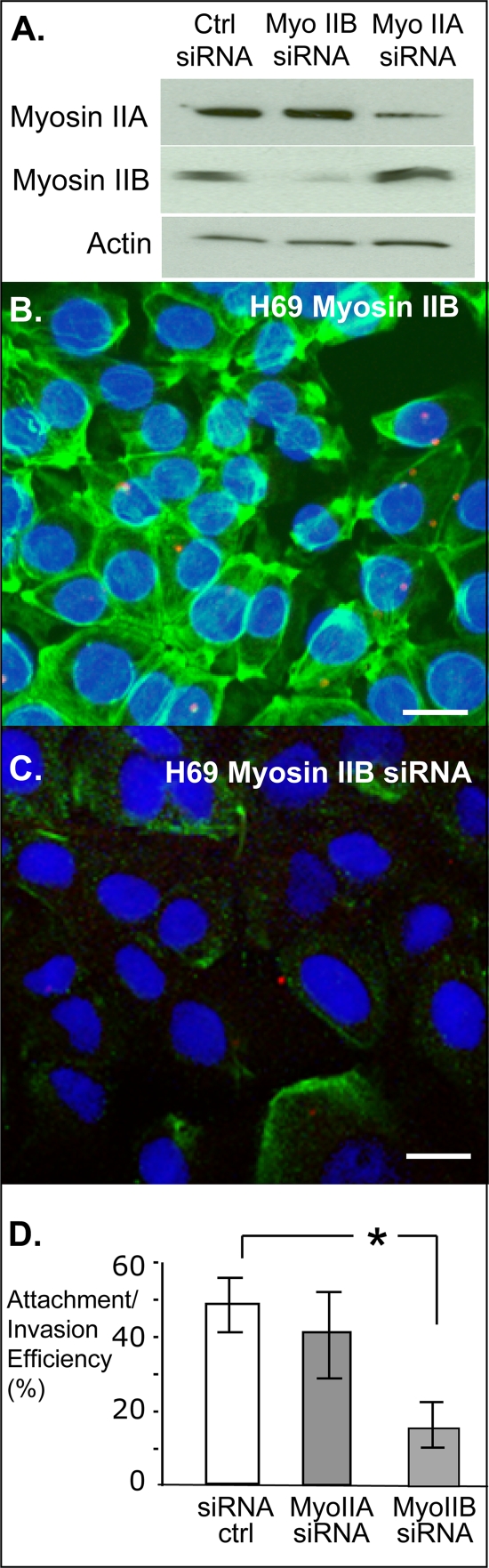
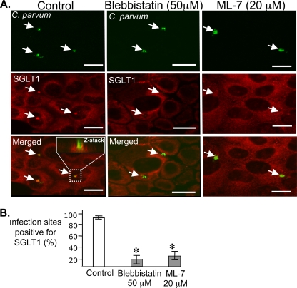
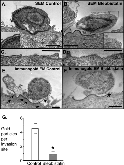
Internalization of the obligate intracellular apicomplexan parasite, Cryptosporidium parvum, results in the formation of a unique intramembranous yet extracytoplasmic niche on the apical surfaces of host epithelial cells, a process that depends on host cell membrane extension. We previously demonstrated that efficient C. parvum invasion of biliary epithelial cells (cholangiocytes) requires host cell actin polymerization and localized membrane translocation/insertion of Na(+)/glucose cotransporter 1 (SGLT1) and of aquaporin 1 (Aqp1), a water channel, at the attachment site. The resultant localized water influx facilitates parasite cellular invasion by promoting host-cell membrane protrusion. However, the molecular mechanisms by which C. parvum induces membrane translocation/insertion of SGLT1/Aqp1 are obscure. We report here that cultured human cholangiocytes express several nonmuscle myosins, including myosins IIA and IIB. Moreover, C. parvum infection of cultured cholangiocytes results in the localized selective aggregation of myosin IIB but not myosin IIA at the region of parasite attachment, as assessed by dual-label immunofluorescence confocal microscopy. Concordantly, treatment of cells with the myosin light chain kinase inhibitor ML-7 or the myosin II-specific inhibitor blebbistatin or selective RNA-mediated repression of myosin IIB significantly inhibits (P < 0.05) C. parvum cellular invasion (by 60 to 80%). Furthermore ML-7 and blebbistatin significantly decrease (P < 0.02) C. parvum-induced accumulation of SGLT1 at infection sites (by approximately 80%). Thus, localized actomyosin-dependent membrane translocation of transporters/channels initiated by C. parvum is essential for membrane extension and parasite internalization, a phenomenon that may also be relevant to the mechanisms of cell membrane protrusion in general.


Figures

FIG. 1.
FIG. 1.
Immunofluorescence C. parvum invasion assay. (A) Cells pretreated with DMSO vehicle (control) are readily infected with C. parvum sporozoites, as detected with immunofluorescence. Pretreatment of cholangiocytes with the myosin II inhibitor blebbistatin (B) or with the myosin light chain kinase inhibitor ML-7 (C) diminishes the number of C. parvum invasion sites detected with immunofluorescence. (D) Quantitation of attachment and attachment/invasion shows that pretreatment of cells with blebbistatin does not affect parasite attachment to H69 cells but that ML-7 reduces attachment approximately 40%. Conversely, blebbistatin pretreatment results in a 10-fold decrease in cellular invasion while ML-7 results in a >2-fold decrease in cellular invasion. Bars = 20 μm. Data are presented as means ± SE. *, P < 0.01 for comparison to control, vehicle-treated cells by ANOVA.
FIG. 2.
FIG. 2.
Effects of the myosin II-specific inhibitor blebbistatin on sporozoite morphology and motility. (A) Sporozoite morphology after treatment with 50 μM blebbistatin or 10 mM 2,3-BDM, a broad inhibitor of myosin ATPase activity. The parasites maintain an elongated phenotype in the presence of blebbistatin yet appear rounded with blunt ends following 2,3-BDM treatment. (B) Quantitation of sporozoite length-to-width ratio. Blebbistatin had no effect on parasite morphology while 2,3-BDM dramatically altered C. parvum shape. (C) Immunofluorescence-based C. parvum motility assay with antigenic trails indicative of gliding cell motility. The parasites exhibited gliding cell motility when treated with 50 μM blebbistatin while this mode of motility was inhibited in the presence of 2,3-BDM. (D) Quantitation of motility trail length. Motility trail length was quantified using Adobe Photoshop software. Data are presented as means ± SE. *, P < 0.01 for comparison to control, vehicle-treated cells by ANOVA.
FIG. 3.
FIG. 3.
H69 cells express myosins IIA and IIB. (A) RT-PCR (RT) was performed for myosin IIA-C on H69 and control HT-29 cells. Both myosin IIA and myosin IIB were detected in H69 cells, while all myosin II isoforms (IIA-C) were detected in control HT29 cells. RT-PCR for 18S was performed on both H69 and HT29 to verify the integrity of the cDNA, and negative controls lacking cDNA (neg) were performed for each primer pair. (B) Immunofluorescence localization of myosins IIA (red) and IIB (green) in subconfluent H69 cells reveals distinct localization patterns. Both IIA and IIB localize to the cell periphery (yellow), while myosin IIB is also distributed throughout the cell. Bar = 20 μm.
FIG. 4.
FIG. 4.
Myosin IIB localizes to sites of C. parvum invasion in confluent cells. (A) Confocal microscopy with dual labeling of nuclei (DAPI; blue) and myosin IIA (green) demonstrates that this isoform localizes primarily to stress fibers throughout C. parvum-infected cells. Arrowheads indicate C. parvum invasion sites identified by DAPI nuclear stain. (B) The blue channel (DAPI staining) was digitally removed to reveal myosin IIA immunofluorescence. No myosin IIA was detected at any infection sites. (C) Confocal microscopy of C. parvum (C.p.; red)-infected confluent H69 cells reveals localized accumulation of myosin IIB (green) to sites of invasion. (D) Both blue (DAPI) and red (C. parvum) channels were removed to reveal localized accumulation of myosin IIB. Bars = 10 μm.
FIG. 5.
FIG. 5.
Phospho-MLC accumulates at C. parvum invasion sites. (A) Confocal immunofluorescence was utilized to assess the distribution of phosphorylated MLC in C. parvum (red)-infected cells. (B) Nearly every infection site showed a strong colocalization of phospho-MLC (green). (C) Merged image of panels A and B demonstrates colocalization of C. parvum and phosphorylated myosin light chain. Phosphorylated myosin light chain aggregates at a region directly adjacent to the invading parasite as seen in the Z section. (D) An immunoblot using a phospho-MLC specific antibody detects increased phoshorylated myosin light chain in cultured cells following C. parvum infection. Actin was blotted as a loading control. Bars = 20 μm.
FIG. 6.
FIG. 6.
Selective RNAi-mediated myosin IIB knockdown diminishes C. parvum cellular invasion. (A) Immunoblots were used to demonstrate selective RNAi-mediated knockdown of myosins IIA and IIB. An siRNA directed against myosin IIA diminishes myosin IIA protein expression, while myosin IIB expression is unaffected. Conversely, a siRNA directed against myosin IIB selectively knocks down this protein. A nonspecific siRNA (no sequence similarity to known genes observed with a BLAST search) was used as a control. (B) Confocal immunofluorescence of C. parvum-infected, control siRNA-treated H69 cells. These cells exhibit robust myosin IIB expression (green) and are readily infected with C. parvum (red); nuclei were stained with DAPI (blue). (C) Confocal immunofluorescence of C. parvum-infected, myosin IIB siRNA-treated H69 cells. These cells exhibit diminished myosin IIB expression, and fewer C. parvum infection sites were detected. (D) Quantitation of attachment/invasion efficiency (number of parasites/100 cells) from siRNA-treated control cells, myosin IIA siRNA-treated cells, and myosin IIB siRNA-treated cells. Bars = 20 μm. Data are presented as means ± SE. *, P < 0.05 for comparison to control siRNA-treated cells by ANOVA.
FIG. 7.
FIG. 7.
Blebbistatin inhibits SGLT1 accumulation at C. parvum invasion sites. (A) Representative confocal micrographs demonstrate that SGLT1 accumulates to regions of C. parvum invasion in control, vehicle-treated cells (left column), while pretreatment of H69 cells with blebbistatin (center column) or ML-7 (right column) inhibits C. parvum-induced aggregation of SGLT1. The top row shows representative images of C. parvum-specific immunofluorescence invasion sites (green), the middle row shows SGLT1-specific immunofluorescence (red), and the bottom row shows the merged images of C. parvum and SGLT1 immunofluorescence. The inset shows a representative confocal x-z plane analysis of the respective boxed area. The x-z plane analysis demonstrates the localized accumulation of SGLT1 in control cells. Bars = 20 μm. (B) Quantitation of SGLT1 aggregation to infection sites from control, blebbistatin-treated, and ML-7-treated cells. Data are presented as means ± SE. *, P < 0.01 for comparison to control, vehicle-treated cells by ANOVA.
FIG. 8.
FIG. 8.
Electron microscopic analysis of invasion sites. (A) Scanning electron micrograph (SEM) from a representative invasion site in control, vehicle-treated H69 cells, showing a fully internalized parasite on the surface of the cell. The parasitophorous vacuole membrane consists of a typical double membrane structure (inset). (B) Scanning electron micrograph (SEM) from representative invasion site in blebbistatin-pretreated cells. The typical parasitophorous vacuole membrane is lacking; rather, a single, discontinuous membrane is present (inset). Over 25 electron micrographs were analyzed for both conditions. Seventy-six percent of the invasion sites from control cells were fully internalized and exhibited the bimembrane parasitophorous vacuole membrane, while fewer than 10 percent of the blebbistatin-treated cells were fully internalized in a bimembrane parasitophorous vacuole membrane. The dense band formed in approximately 50% of invasion sites in both control and blebbistatin-treated cells. (C, D) Representative images of the plasma membrane of control (C) and blebbistatin-treated, uninfected (D) cells. No obvious ultrastructural morphological differences were noted in this region. (E) Representative Immunogold electron micrograph (EM) detecting SGLT1 at C. parvum invasion sites in control, vehicle-treated cells. Gold particles localized to the host cell parasite interface (arrows), while few gold particles were detected at C. parvum invasion sites in blebbistatin-pretreated cells (F). (G) Quantitation of nanogold particles localized to invasion sites in control cells and blebbistatin-treated cells. Bars = 0.5 μm. Data are presented as means ± SE. *, P < 0.02 for comparison to control, vehicle-treated cells by Student's t test.

Similar articles

Cited by

References

    1. Agre, P., L. S. King, M. Yasui, W. B. Guggino, O. P. Ottersen, Y. Fujiyoshi, A. Engel, and S. Nielsen. 2002. Aquaporin water channels—from atomic structure to clinical medicine. J. Physiol. 542:3-16. - PMC - PubMed
    1. Aji, T., T. Flanigan, R. Marshall, C. Kaetzel, and M. Aikawa. 1991. Ultrastructural study of asexual development of Cryptosporidium parvum in a human intestinal cell line. J. Protozool. 38:82S-84S. - PubMed
    1. Bao, J., S. S. Jana, and R. S. Adelstein. 2005. Vertebrate nonmuscle myosin II isoforms rescue small interfering RNA-induced defects in COS-7 cell cytokinesis. J. Biol. Chem. 280:19594-19599. - PubMed
    1. Bao, J., X. Ma, C. Liu, and R. S. Adelstein. 2007. Replacement of nonmuscle myosin II-B with II-A rescues brain but not cardiac defects in mice. J. Biol. Chem. 282:22102-22111. - PubMed
    1. Bonnin, A., A. Lapillonne, T. Petrella, J. Lopez, C. Chaponnier, G. Gabbiani, S. Robine, and J. F. Dubremetz. 1999. Immunodetection of the microvillous cytoskeleton molecules villin and ezrin in the parasitophorous vacuole wall of Cryptosporidium parvum (Protozoa: Apicomplexa). Eur. J. Cell Biol. 78:794-801. - PubMed

Publication types

MeSH terms