. 2010 Jul;153(3):1016-30.
doi: 10.1104/pp.110.158048. Epub 2010 May 10.

The acidic A-domain of Arabidopsis TOC159 occurs as a hyperphosphorylated protein

Affiliations

The acidic A-domain of Arabidopsis TOC159 occurs as a hyperphosphorylated protein

Birgit Agne et al. Plant Physiol. 2010 Jul.

Abstract

The translocon at the outer membrane of the chloroplast assists the import of a large class of preproteins with amino-terminal transit sequences. The preprotein receptors Toc159 and Toc33 in Arabidopsis (Arabidopsis thaliana) are specific for the accumulation of abundant photosynthetic proteins. The receptors are homologous GTPases known to be regulated by phosphorylation within their GTP-binding domains. In addition to the central GTP-binding domain, Toc159 has an acidic N-terminal domain (A-domain) and a C-terminal membrane-anchoring domain (M-domain). The A-domain of Toc159 is dispensable for its in vivo activity in Arabidopsis and prone to degradation in pea (Pisum sativum). Therefore, it has been suggested to have a regulatory function. Here, we show that in Arabidopsis, the A-domain is not simply degraded but that it accumulates as a soluble, phosphorylated protein separated from Toc159. However, the physiological relevance of this process is unclear. The data show that the A-domain of Toc159 as well as those of its homologs Toc132 and Toc120 are targets of a casein kinase 2-like activity.


Figures

Figure 1.
Figure 1.
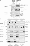
Transgenic plants for TAP of AtToc159. A, Schematic representation of the Toc159-TAP fusion proteins. *, In the C-terminally fused TAP tag, the order of its segments is inversed and reads CBP-TEV-ProAProA. B, Immunoblot of plant extracts of transgenic lines with unspecific rabbit IgG to detect the TAP-Toc159 fusion proteins (lanes 3–5) or the TAP tag alone (lane 7). Anti-Toc159A antibodies were used to detect endogenous AtToc159 in wild-type (WT) plants (lane 1). C, TAP-Toc159:ppi2 and Toc159A-TAP:WT plants reveal a green phenotype. Seedlings were grown for 21 d under short-day conditions on medium containing phosphinotricin before the photographs were taken. Bars = 0.2 cm.
Figure 2.
Figure 2.
IgG affinity purification of TAP-Toc159 species from soluble and membrane fractions. A, Immunoblot analysis of total protein extracts (EXTR), soluble fraction (SN100), and membrane fraction (P100) of wild-type plants expressing only the TAP tag (lanes 1–3) and ppi2 plants expressing TAP-Toc159 (lanes 4–6). Fifty micrograms of protein from each fraction was separated by SDS-PAGE and subjected to western-blot analysis with antibodies as indicated. B, Immunoblot analysis of different fractions obtained during purification of the TAP tag/TAP-Toc159 from the soluble and membrane fractions. Fifty milligrams of soluble protein (SN100) and 20 mg of Triton X-100-solubilized membrane-associated protein (P100) were loaded on an IgG Affi-Gel column. Proteins were eluted using TEV protease. The percentage indicates the amount of protein loaded relative to the total amount of protein in the load, unbound and eluate fractions (100%). LHCP, Chlorophyll a/b-binding protein; LSU, large Rubisco subunit; PRK, phosphoribulokinase; TAP, samples originating from TAP:WT plants; 159, samples originating from TAP-Toc159:ppi2 plants; TAP-Toc159*/Toc159*, proteolytic fragments of TAP-Toc159/Toc159.
Figure 3.
Figure 3.
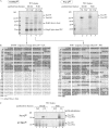
The Toc159 species purified from the soluble fraction constitute the A-domain. A, TEV eluates after purification of TAP-Toc159 from TAP-Toc159:ppi2 (159) and of the control TAP tag from TAP:WT tag plants (TAP) were separated on a NuPAGE Novex 4% to 12% BisTris gel. The gel was stained with the phosphospecific fluorescent dye ProQ-Diamond and with the total protein stain SyproRuby. The PeppermintStick molecular mass marker (Invitrogen) contains two phosphorylated proteins of 45 kD (ovalbumin) and 24 kD (β-casein). Toc159*, Proteolytic fragment of Toc159. B, Peptides (boldface and underlined) and sequence coverage obtained by tandem mass spectrometry analysis of TAP-Toc159 species. The Toc159 A-domain sequence, inferred from the experimentally determined start of the Toc86 fragment of pea Toc159 (Hirsch et al., 1994; Bolter et al., 1998), is highlighted in gray. C, Phosphatase treatment demonstrates phosphospecific staining of Toc159 species. TEV eluates derived from TAP:WT tag plants (TAP) or TAP-Toc159:ppi2 plants (159) were treated (+) or not (−) with λ-phosphatase prior to separation on a NuPAGE Novex 4% to 12% BisTris gel and ProQ-Diamond and SyproRuby staining. SN100, Protein purified from the soluble fraction; P100, protein purified from the 100,000g pellet solubilized with Triton X-100.
Figure 4.
Figure 4.
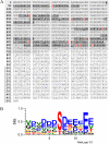
Toc159 phosphopeptides map to the A-domain. A, Sequence of AtToc159 with phosphopeptides (boldface and underlined) and phosphorylation sites (red) according to Table I. B, WebLogo analysis (Crooks et al., 2004) of Toc159 phosphorylation sites. Color scheme is as follows: S/T, red; D/E, blue; K/R, orange; L/I/V/M, green.
Figure 5.
Figure 5.
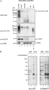
Toc159A overexpressed in planta is phosphorylated. A, Immunoblot analysis of different fractions obtained during purification of Toc159A(aa1-740)-TAP from the soluble fraction (SN100). Soluble protein (lanes 3 and 4) was loaded on a HsIgG-Affi-Gel column. After removal of unbound protein (UB; lane 5) and washing, elution was carried out using AcTEV protease (TEV; lane 6). EXTR, Plant extract; P100, 100,000g pellet; SN100, 100,000g supernatant; L, 100,000g supernatant at the time of loading on HsIgG-Affi-Gel. B, The PeppermintStick phosphoprotein molecular mass standard (MW) and the TEV eluate derived from the soluble fraction of WT:Toc159A-TAP plants (159A) were treated (+) or not (−) with λ-phosphatase prior to separation on a NuPAGE Novex 4% to 12% BisTris gel and ProQ-Diamond and SyproRuby staining. Asterisks indicate phosphorylated proteins of the molecular mass standard.
Figure 6.
Figure 6.
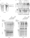
In vitro phosphorylation of Toc159A by an Arabidopsis kinase activity sensitive to heparin and using GTP. A, Toc159A-His-6x (Toc159aa1-740-His-6x) was overexpressed in E. coli BL21(DE3) and purified under native conditions from the bacterial soluble protein fraction using nickel-nitrilotriacetic acid agarose affinity chromatography. Samples of noninduced cultures (−IPTG [isopropylthio-β-galactoside]), induced cultures (+IPTG), the soluble protein fraction (soluble), and the purified protein (purif.) were separated by SDS-PAGE followed by Coomassie Brilliant Blue staining. B, Bovine serum albumin (BSA; control) or purified Toc159A-His-6x was incubated in the presence of [γ-33P]ATP with different amounts of Arabidopsis plant cell extract (Total; lanes 2, 6, and 7), the supernatant (SN100; lanes 3, 8, and 9), or the pellet (P100; lanes 4, 10, and 11) fraction after 100,000g centrifugation of the extract. Incubation in the absence of plant protein (lanes 1 and 5) was used to monitor autophosphorylation. The samples were analyzed by SDS-PAGE followed by Coomassie Brilliant Blue staining (bottom) and phosphorimager analysis (top). To ease detection of phosphorylated A-domain fragments in the plant samples, the Mr values of the characteristic fragments of Toc159A-His-6x, observed after bacterial overexpression and purification, are marked by arrows. C, In vitro phosphorylation of Toc159A-His-6x by the SN100 (left) and P100 (right) fractions in the presence of the kinase inhibitors heparin, DRB, chelerythrine chloride, and apigenin or unlabeled GTP as an alternative phosphate donor.
Figure 7.
Figure 7.
Phosphorylation of recombinant A-domains by CK2. A, One microgram of purified Toc159A-His-6x or casein as a control was incubated without (lanes 1 and 4) or with (lanes 2 and 5) recombinant maize CK2 α-subunit and ATP/[γ-33P]ATP as the phosphate donor. In addition, phosphorylation of Toc159A-His-6x with CK2 was performed in the presence of 15 μg mL−1 heparin as an inhibitor (lane 3). The samples were separated by SDS-PAGE followed by Coomassie Brilliant Blue staining (lanes 1′–5′) and phosphorimager analysis (lanes 1–5). B, One microgram of purified Toc33G-His-6x, Toc120A-His-6x, Toc132A-His-6x, Toc159A-His-6x, or GST-Toc159G was incubated without (even lanes) or with (odd lanes) recombinant CK2 and ATP/[γ-33P]ATP as the phosphate donor. The samples were separated by SDS-PAGE followed by Coomassie Brilliant Blue staining (lanes 1′–10′) and phosphorimager analysis (lanes 1–10).
Figure 8.
Figure 8.
Inhibition of in vitro chloroplast protein import by heparin. A, Isolated Arabidopsis chloroplasts were preincubated without or with 15 μg mL−1 heparin, 6 μm DRB, 3 units μL−1 recombinant maize CK2 α-subunit, 50 μm apigenin, or 10 mm glycerol-2-phosphate for 20 min at 25°C in the dark. Then, in vitro-translated, [35S]Met-labeled preprotein of the small subunit of Rubisco (pSSu) was added and import was allowed to proceed for 0, 7.5, and 15 min. B, Quantification of the effect of heparin on chloroplast protein import. The graph shows the quantification of the amount of imported SSu at 15 min in three independent experiments. In both panels, the amount of SSu imported into wild-type (WT) chloroplasts without the addition of inhibitor at 15 min was set to 100%. IVT, In vitro translate; M, molecular mass standard. Asterisks indicate pSSu modified in the course of the import reactions.
Figure 9.
Figure 9.
Alignment of the junction region between A- and G-domains of the Toc159 proteins of Oryza sativa (Os), Vitis vinifera (Vv), Populus trichocarpa (Pt), Pisum sativum (Ps), and Arabidopsis thaliana (At). Tryptic peptides identified in the soluble (SN100) or membrane (P100) fraction and the N terminus of pea Toc86 are shown. In addition, the positions of a semitryptic peptide identified in the soluble fraction and the region where Toc159 processing is likely to occur are shown.

Similar articles

Cited by

References

    1. Agne B, Infanger S, Wang F, Hofstetter V, Rahim G, Martin M, Lee DW, Hwang I, Schnell D, Kessler F. (2009) A toc159 import receptor mutant, defective in hydrolysis of GTP, supports preprotein import into chloroplasts. J Biol Chem 284: 8670–8679 - PMC - PubMed
    1. Aronsson H, Combe J, Patel R, Jarvis P. (2006) In vivo assessment of the significance of phosphorylation of the Arabidopsis chloroplast protein import receptor, atToc33. FEBS Lett 580: 649–655 - PubMed
    1. Bauer J, Chen K, Hiltbunner A, Wehrli E, Eugster M, Schnell D, Kessler F. (2000) The major protein import receptor of plastids is essential for chloroplast biogenesis. Nature 403: 203–207 - PubMed
    1. Blom N, Sicheritz-Ponten T, Gupta R, Gammeltoft S, Brunak S. (2004) Prediction of post-translational glycosylation and phosphorylation of proteins from the amino acid sequence. Proteomics 4: 1633–1649 - PubMed
    1. Bolter B, May T, Soll J. (1998) A protein import receptor in pea chloroplasts, Toc86, is only a proteolytic fragment of a larger polypeptide. FEBS Lett 441: 59–62 - PubMed

Publication types

MeSH terms

LinkOut - more resources