. 2010 Jul;30(14):3672-84.
doi: 10.1128/MCB.00223-10. Epub 2010 May 10.

The POU transcription factor Drifter/Ventral veinless regulates expression of Drosophila immune defense genes

Affiliations

The POU transcription factor Drifter/Ventral veinless regulates expression of Drosophila immune defense genes

Anna Junell et al. Mol Cell Biol. 2010 Jul.

Abstract

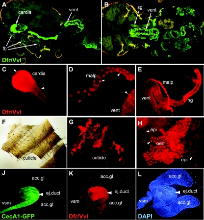
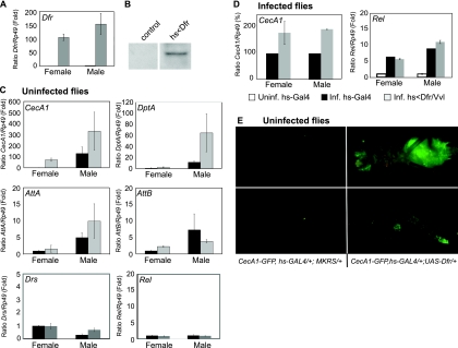
Innate immunity operates as a first line of defense in multicellular organisms against infections caused by different classes of microorganisms. Antimicrobial peptides (AMPs) are synthesized constitutively in barrier epithelia to protect against microbial attack and are also upregulated in response to infection. Here, we implicate Drifter/Ventral veinless (Dfr/Vvl), a class III POU domain transcription factor, in tissue-specific regulation of the innate immune defense of Drosophila. We show that Dfr/Vvl is highly expressed in a range of immunocompetent tissues, including the male ejaculatory duct, where its presence overlaps with and drives the expression of cecropin, a potent broad-spectrum AMP. Dfr/Vvl overexpression activates transcription of several AMP genes in uninfected flies in a Toll pathway- and Imd pathway-independent manner. Dfr/Vvl activates a CecA1 reporter gene both in vitro and in vivo by binding to an upstream enhancer specific for the male ejaculatory duct. Further, Dfr/Vvl and the homeodomain protein Caudal (Cad) activate transcription synergistically via this enhancer. We propose that the POU protein Dfr/Vvl acts together with other regulators in a combinatorial manner to control constitutive AMP gene expression in a gene-, tissue-, and sex-specific manner, thus promoting a first-line defense against infection in tissues that are readily exposed to pathogens.


Figures

FIG. 1.
FIG. 1.
Dfr/Vvl is localized to the nuclei of fat body, digestive system, and barrier epithelia in uninfected larvae. Shown are immunostaining of Dfr/Vvl (red) in dissected tissues from third instar larvae and staining of nuclei with DAPI (4′,6-diamidino-2-phenylindole) (blue). Strong nuclear Dfr/Vvl staining was evident in the fat body (fb) (A and A′), salivary glands (sg) (B and B′), and trachea (tr) (C and C′). In the malpighian tubules (malp), different regions reproducibly conferred either equal distributions between the nuclei and cytoplasm (D and D′) or predominantly nuclear distribution (E and E′). In the digestive system, Dfr/Vvl staining was intense in the proventriculus (pv) (F, G, and G′), in gastric ceca (gc) (F), and in the hindgut (hg) (H and H′). In the midgut (mg), Dfr/Vvl staining was weak and diffuse (F and H). Also shown are differential interference contrast micrograph of the larval cuticle (cut) and epidermis (epi), with two ventral denticle belts (db) visible (J) and fluorescence micrographs of the same specimen showing nuclear localization of Dfr/Vvl (J′) and DAPI (J") in epidermal cells. Tissues/organs were named according to M. Demerec (45).
FIG. 2.
FIG. 2.
Dfr/Vvl is expressed in immunocompetent barrier epithelia of uninfected flies. Frozen cryostat sections of the thorax and abdomen of a fly in sagittal (A) and horizontal (B) sections revealed strong Dfr/Vvl immunostaining (green) in the digestive tract, especially in the cardia, ventriculus (vent), and salivary glands (sg). In addition, Dfr/Vvl was apparent in the fat body (fb). Anterior is to the left. (C to E) Dissected organs from adults confirmed strong Dfr/Vvl immunoreactivity (red) in the digestive system, in the cardia, malpighian tubules (malp), ventriculus (vent), and hindgut (hg). Dissected cuticle of a male fly in bright field (F) and in epifluorescence (G and H) revealed intense nuclear Dfr/Vvl immunostaining in oenocytes lying just beneath the cuticle (G) and in higher magnification (H). (H) Weak staining in nuclei of epidermal cells (epi) was also evident. Arrowheads point to nuclear Dfr/Vvl staining. Dissected male reproductive organs from an uninfected fly show the distribution of CecA1-GFP (J), the Dfr/Vvl protein (K), and nuclear DAPI (L) staining. Expression was strong in the ejaculatory duct (ej.duct) but absent from the accessory glands (acc.gl.), seminal vesicle (vsm), and testis (not shown).
FIG. 3.
FIG. 3.
Constitutive CecA1 expression in the ejaculatory duct is Toll pathway and Imd pathway independent. Constitutive expression of CecA1-GFP in the ejaculatory duct of uninfected wt (A), Myd88 mutant (B), and imd mutant (C) male flies corresponds to nuclear Dfr/Vvl staining (A′, B′, and C′) and nuclear DAPI staining (A", B", and C").
FIG. 4.
FIG. 4.
Overexpression of Dfr/Vvl-induced expression of AMP genes in an infection-independent manner. (A) Quantification of Dfr/Vvl mRNA by RT-qPCR in Dfr/Vvl-overexpressing flies (hs-GAL4/UAS-Dfrwt) (gray bars) compared to control flies (hs-GAL4/+) (black bars). The level of Dfr/Vvl mRNA in uninfected female control flies was set to 1, using Rp49 levels as an internal control. (B) Western blot analysis of nuclear extracts from control and Dfr/Vvl-overexpressing flies, with genotypes as described in the legend for panel A. (C) RT-qPCR analysis of AMP gene expression in uninfected Dfr/Vvl-overexpressing flies (hs-GAL4/UAS-Dfrwt) (gray bars) compared to control flies (hs-GAL4+>) (black bars). The relative mRNA levels of each gene (CecA1, Dpt, AttA, AttB, Drs, and Rel) in extracts of female or male flies of each genotype were calculated relative to the expression of the same gene in female control flies, which was set to 1, using Rp49 mRNA as an internal control. (D) RT-qPCR analysis of CecA1 and Rel levels in infected females and males overexpressing Dfr/Vvl (hs-GAL4/UAS-Dfrwt) (gray bars) compared to control (hs-GAL4/+) flies (black bars). The level of CecA1 mRNA in infected control flies was set to 100%. The level of Rel mRNA in infected flies was calculated relative to the expression of the same gene in uninfected female control (hs-GAL4/+) flies, set to 1. (E) Ubiquitous overexpression of Dfr/Vvl (hs-GAL4, CecA1-GFP; UAS-Dfrwt/+) by heat shock treatment promoted CecA1-GFP expression in the fat body of the head, thorax, and abdomen (top right panel) and in several regions of the midgut (bottom right panel) of uninfected flies. In control flies (hs-GAL4, CecA1-GFP; +/+) (left panels), no CecA1-GFP expression was induced by the heat shock.
FIG. 5.
FIG. 5.
Identification of a Dfr/Vvl-dependent CecA1 regulatory region. (A to D) Constitutive CecA1 expression in male reproductive organs in transgenic flies carrying different CecA1-lacZ constructs. Reporter β-galactosidase staining was specifically localized to the ejaculatory duct (ej.duct) but was absent from the accessory glands (acc.glands), seminal vesicle (vsm), and testis. Expression was evident from constructs carrying 760 bp upstream of the CecA1 transcription start site (21) (A, B, and C), even if the regulatory R1 and κB sequences had been deleted (55) (B) or the GATA site mutated (53) (C). A construct containing 111 bp of the CecA1 upstream sequence did not confer any β-Gal staining in the ejaculatory duct (D), indicating that necessary elements must be located in the bp −760 to −111 region. (E) Quantitative measurements of luciferase activity in extracts of mbn-2 cells after expression of Dfr/Vvl, Cad, or Dfr/Vvl and Cad proteins, with a panel of CecA1-luc reporter constructs, carrying progressive 5′ deletions (top graph) or specific mutations in putative Dfr/Vvl binding sites (bottom graph), as indicated. The upstream region and 5′ untranslated region (UTR) of the CecA1 gene (bp −751 to +71) is represented by a horizontal line. The EMSA control probe (C) and the location of the proximal regulatory region, containing the R1, κB, and GATA motifs, are indicated (not to scale). The extent of the deletion in CecA1Δ-luc (bp −466 to −403) is indicated by Δ and by parentheses in the sequences below. The sequences of probes E1 to E3 used in the EMSA are shown (note that each overlaps with the previous probe by 5 bp). The putative Dfr/Vvl binding sites are shown in red, and mutated base pairs in the CecA1-luc constructs are underlined. The results are presented as the mean values of at least three independent experiments (± standard error of the mean [SEM]). (F) EMSA with purified, GST-tagged Dfr/Vvl, and GST protein alone, using four different 32P-labeled CecA1 probes, E1 (bp −493 to −454), E2 (bp −458 to −419), E3 (bp −423 to −380) covering partly overlapping parts of the bp −484 to −377 region 5′ of CecA1, and, as a control, an upstream fragment (C; bp −663 to −644). GST-tagged Dfr/Vvl protein, but not GST alone, bound with high affinity to the E1 and E3 probes, while only weak binding was observed with the E2 probe. Dfr/Vvl protein did not bind to the control (C) fragment (bp −663 to −644), and no binding was observed for lanes without any protein added (−).
FIG. 6.
FIG. 6.
An upstream enhancer regulates constitutive, tissue-specific expression of CecA1. Shown are a schematic representation of the genomic region upstream of the CecA1 coding sequence and a model for the organization of cis-regulatory elements present in this region. A tissue-specific enhancer that directs expression in the ejaculatory duct, named the male ejaculatory duct enhancer, is proposed to be targeted by Dfr/Vvl (stars) and Cad (hexagons), each binding to different sequence elements (E1 to E3) within the enhancer. Binding of Dfr/Vvl to the CANTAAA sequence motif in the enhancer activates expression of CecA1. The proximal regulatory region (gray box), which is a target of infection-induced signaling and binding of Rel and GATA factors (gray dimer), does not appear to be required for the constitutive expression in the ejaculatory duct, since CecA1-lacZ constructs with deletions/mutations in this region conferred ejaculatory duct-specific reporter gene expression. The model is based on results from this and previous studies (21, 29, 53, 55, 59, 70, 75).

Similar articles

Cited by

References

    1. Aerts, A. M., I. E. Francois, B. P. Cammue, and K. Thevissen. 2008. The mode of antifungal action of plant, insect and human defensins. Cell. Mol. Life Sci. 65:2069-2079. - PMC - PubMed
    1. Agerberth, B., and G. H. Gudmundsson. 2006. Host antimicrobial defence peptides in human disease. Curr. Top. Microbiol. Immunol. 306:67-90. - PubMed
    1. Akira, S., S. Uematsu, and O. Takeuchi. 2006. Pathogen recognition and innate immunity. Cell 124:783-801. - PubMed
    1. Andersen, B., and M. G. Rosenfeld. 2001. POU domain factors in the neuroendocrine system: lessons from developmental biology provide insights into human disease. Endocr. Rev. 22:2-35. - PubMed
    1. Anderson, M. G., S. J. Certel, K. Certel, T. Lee, D. J. Montell, and W. A. Johnson. 1996. Function of the Drosophila POU domain transcription factor drifter as an upstream regulator of breathless receptor tyrosine kinase expression in developing trachea. Development 122:4169-4178. - PubMed

Publication types

MeSH terms

LinkOut - more resources