. 2010 Jun;298(6):C1424-30.
doi: 10.1152/ajpcell.00503.2009. Epub 2010 Mar 24.

A mechanosensitive ion channel regulating cell volume

Affiliations

A mechanosensitive ion channel regulating cell volume

Susan Z Hua et al. Am J Physiol Cell Physiol. 2010 Jun.

Abstract

Cells respond to a hyposmotic challenge by swelling and then returning toward the resting volume, a process known as the regulatory volume decrease or RVD. The sensors for this process have been proposed to include cationic mechanosensitive ion channels that are opened by membrane tension. We tested this hypothesis using a microfluidic device to measure cell volume and the peptide GsMTx4, a specific inhibitor of cationic mechanosensitive channels. GsMTx4 had no effect on RVD in primary rat astrocytes or Madin-Darby canine kidney (MDCK) cells but was able to completely inhibit RVD and the associated Ca(2+) uptake in normal rat kidney (NRK-49F) cells in a dose-dependent manner. Gadolinium (Gd(3+)), a nonspecific blocker of many mechanosensitive channels, inhibited RVD and Ca(2+) uptake in all three cell types, demonstrating the existence of at least two types of volume sensors. Single-channel stretch-activated currents are present in outside-out patches from NRK-49F, MDCK, and astrocytes, and they are reversibly inhibited by GsMTx4. While mechanosensitive channels are involved in volume regulation, their role for volume sensing is specialized. The NRK cells form a stable platform from which to screen drugs that affect volume regulation via mechanosensory channels and as a sensitive system to clone the channel.


Figures

Fig. 1.
Fig. 1.
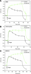
Effects of GsMTx4 on cell volume regulation of normal rat kidney (NRK-49F) cells (A and B), primary rat astrocytes (C), and Madin-Darby canine kidney (MDCK) cells (D). The chamber was perfused with isotonic solution (Iso) for 10–15 min and then switched to hypotonic solution (Hypo) at the times indicated. The ratio of the volume change to the resting cell volume, ΔV/Vo, is plotted in AD. Both GsMTx4 L enantiomer (5 μM) and D enantiomer (2.5 μM) blocked regulatory volume decrease (RVD) in the NRK cells (control and L-form, n = 5; D-form, n = 3). The cells responded in a dose-dependent fashion (B) with an apparent Kd of 100–200 nM. In primary rat astrocytes (n = 5) and MDCK (n = 6) cells, 5 μM GsMTx4 in both isotonic and hypotonic solutions had no significant effect on RVD (C and D).
Fig. 2.
Fig. 2.
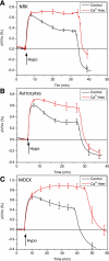
Sensitivity of RVD to Gd3+ for NRK-49F (A), primary rat astrocytes (B), and MDCK cells (C). Gd3+ (100 μM) in isotonic and hypotonic solutions inhibited RVD in all three cell types, causing a slow, extended, increase in volume (NRK, n = 4; astrocytes, n = 3; MDCK, n = 3).
Fig. 3.
Fig. 3.
The dependence of cell volume regulation on extracellular Ca2+ for NRK-49F (A), primary rat astrocytes (B), and MDCK cells (C). The removal of Ca2+ suppressed RVD in all three cell types (NRK, n = 4; astrocytes, n = 4; MDCK, n = 3).
Fig. 4.
Fig. 4.
Response of intracellular Ca2+ to hypotonic challenges in NRK-49F cells. A and B: Ca2+ Fluo-4 images before (A) and after (B) hypotonic challenge in control solutions. C: typical traces of the time dependence of the Ca2+ increase from representative cells marked in A and B are plotted as the fractional change in fluorescence (IIo)/Io. Similar responses were observed in ∼80% of cells. DF: application of 5 μM GsMTx4 in both isotonic and hypotonic solutions eliminated the swelling-induced Ca2+ elevation (same conditions as in AC).
Fig. 5.
Fig. 5.
GsMTx4 has little effect on swelling-induced Ca2+ elevation in astrocytes and MDCK cells. A: time dependence of swelling-induced intracellular Ca2+ in astrocytes as taken from representative cells. B: time dependence of swelling-induced Ca2+ elevation in the presence of 5 μM GsMTx4. C and D: swelling-induced Ca2+ elevation in MDCK cells (as per A and B).
Fig. 6.
Fig. 6.
Inhibition of stretch-activated currents by GsMTx4. A and B: GsMTx4 at 3 μM concentration inhibited pressure-dependent channel activity for MDCK cells (A) and NRK cells (B) for outside-out patches. Outside-out patches were formed and channel activity was monitored at −50 mV with the indicated pressure pulse. Inhibition was reversed with washout with bath solution. The data were averaged for 5–10 pulses before, during, and after (washout) the application of the peptide. C: the average response over three patches, using the current amplitude during the pressure pulse.

Similar articles

Cited by

References

    1. Acher R. Water homeostasis in the living: molecular organization, osmoregulatory reflexes and evolution. Ann Endocrinol (Paris) 63: 197–218, 2002 - PubMed
    1. Arniges M, Vazquez E, Fernandez-Fernandez JM, Valverde MA. Swelling-activated Ca2+ entry via TRPV4 channel is defective in cystic fibrosis airway epithelia. J Biol Chem 279: 54062–54068, 2004 - PubMed
    1. Ateya DA, Sachs F, Gottlieb PA, Besch S, Hua SZ. Volume cytometry: microfluidic sensor for high-throughput screening in real time. Anal Chem 77: 1290–1294, 2005 - PubMed
    1. Becker D, Blase C, Bereiter-Hahn J, Jendrach M. TRPV4 exhibits a functional role in cell-volume regulation. J Cell Sci 118: 2435–2440, 2005 - PubMed
    1. Bode F, Sachs F, Franz MR. Tarantula peptide inhibits atrial fibrillation. Nature 409: 35–36, 2001 - PubMed

Publication types

MeSH terms

LinkOut - more resources