. 2010 May;103(5):2737-46.
doi: 10.1152/jn.01112.2009. Epub 2010 Mar 24.

Co-induction of LTP and LTD and its regulation by protein kinases and phosphatases

Affiliations

Co-induction of LTP and LTD and its regulation by protein kinases and phosphatases

Kathryn B Grey et al. J Neurophysiol. 2010 May.

Abstract

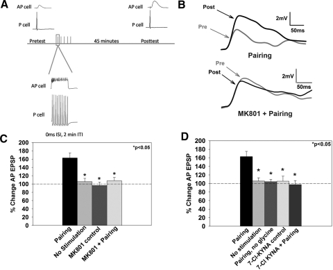
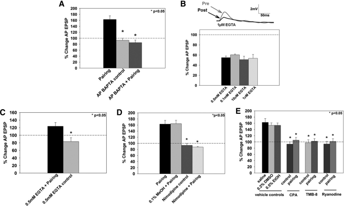
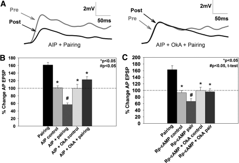
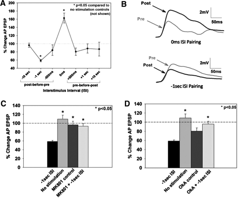
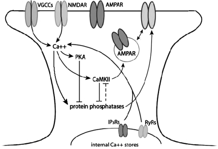
The cellular properties of long-term potentiation (LTP) following pairing of pre- and postsynaptic activity were examined at a known glutamatergic synapse in the leech, specifically between the pressure (P) mechanosensory and anterior pagoda (AP) neurons. Stimulation of the presynaptic P cell (25 Hz) concurrent with a 2 nA depolarization of the postsynaptic AP cell significantly potentiated the P-to-AP excitatory postsynaptic potential (EPSP) in an N-methyl-d-aspartate receptor (NMDAR)-dependent manner based on inhibitory effects of the NMDAR antagonist MK801 and inhibition of the NMDAR glycine binding site by 7-chlorokynurenic acid. LTP was blocked by injection of bis-(o-aminophenoxy)-N,N,N',N'-tetraacetic acid (BAPTA) into the postsynaptic (AP) cell, indicating a requirement for postsynaptic elevation of intracellular Ca(2+). Autocamtide-2-related inhibitory peptide (AIP), a specific inhibitor of Ca(2+)/calmodulin-dependent kinase II (CaMKII), and Rp-cAMP, an inhibitor of protein kinase A (PKA), also blocked pairing-induced potentiation, indicating a requirement for activation of CaMKII and PKA. Interestingly, application of AIP during pairing resulted in significantly depressed synaptic transmission. Co-application of AIP with the protein phosphatase inhibitor okadaic acid restored synaptic transmission to baseline levels, suggesting an interaction between CaMKII and protein phosphatases during induction of activity-dependent synaptic plasticity. When postsynaptic activity preceded presynaptic activity, NMDAR-dependent long-term depression (LTD) was observed that was blocked by okadaic acid. Postsynaptic injection of botulinum toxin blocked P-to-AP potentiation while postsynaptic injection of pep2-SVKI, an inhibitor of AMPA receptor endocytosis, inhibited LTD, supporting the hypothesis that glutamate receptor trafficking contributes to both LTP and LTD at the P-to-AP synapse in the leech.


Figures

Fig. 1.
Fig. 1.
Paired pre- and postsynaptic activity induces potentiation at the pressure (P)-to-anterior pagoda (AP) synapse. A: schematic of the pairing protocol. Pre- and posttest excitatory postsynaptic potential (EPSP) measurements in the postsynaptic AP cell were obtained by eliciting a single presynaptic (P cell) action potential. Pairing consisted of a 10 pulse train (25 Hz, 10 ms) applied to the P-cell coinciding with a 2 nA step depolarization (500 ms) of the AP cell. Posttest EPSP measurements were completed after a 45 min consolidation period. B: representative EPSP traces prior to (pre) and following pairing (post) from synapses that underwent pairing in normal saline (top) or in MK801 (bottom). The gray trace denotes the pretest EPSP and the black trace denotes the posttest EPSP. C: effects of the N-methyl-d-aspartate receptor (NMDAR) antagonist, MK801, on long-term potentiation (LTP). Pairing-induced LTP was blocked by the application of the NMDAR antagonist MK801 (pairing + MK801). No stimulation and MK801 control groups are significantly different from the pairing group, indicating that potentiation only occurs following coordinated activation of the prepostsynaptic neurons. D: role of glycine during LTP. Elimination of glycine from the bath during the training protocol (pairing, no glycine) or application of 7-chlorokynurenic acid (7-Cl KYNA; pairing +7-Cl KYNA), which blocks the NMDAR glycine binding site, prevented pairing-induced potentiation. Administration of 7-Cl KYNA alone (7-Cl-KYNA control) did not significantly affect baseline synaptic transmission. Asterisks indicates statistically significant difference relative to the pairing group.
Fig. 2.
Fig. 2.
Role of multiple sources of postsynaptic Ca2+ during pairing-induced LTP. A: effects of postsynaptic bis-(o-aminophenoxy)-N,N,N′,N′-tetraacetic acid (BAPTA) injection on LTP. No potentiation was observed when BAPTA was injected into the postsynaptic (AP) cell before administration of the pairing protocol (AP BAPTA + pairing). Postsynaptic injection of BAPTA alone did not affect baseline synaptic transmission (AP BAPTA control). B: effects of presynaptic EGTA injection on the P-to-AP EPSP. After 5 min of iontophoretically injected EGTA into the presynaptic (P) cell, evoked P-to-AP synaptic transmission decreased, on average, 50% from preEGTA treatment levels. Top: representative EPSP traces from a P-to-AP synapse treated with 1 μM EGTA. The posttest (post, gray trace) was conducted 1 h after the pretest (pre, black trace). Bottom: averaged change in EPSP size following presynaptic EGTA treatment for concentrations between 1 μM to 0.5 mM. C: effects of presynaptic EGTA injection on LTP. Iontophoresis of 0.5 mM EGTA into the presynaptic (P cell) before administration of the pairing protocol produced potentiation relative to the EGTA control (EGTA treatment, but no pairing). D: effects of nimodipine of LTP. Nimodipine blocked pairing-induced potentiation (nimodipine + pairing). Methanol, the vehicle control for nimodipine, did not affect pairing-induced potentiation (0.1% vol/vol methanol + pairing), and nimodipine administration alone did not affect baseline synaptic transmission (nimodipine control). E: effects of inhibition of release from intracellular Ca2+ stores on LTP. Inhibition of Ca2+ release from intracellular stores also contributed to pairing-induced LTP, which was blocked by depletion of Ca2+ stores by cyclopiazonic acid (CPA), inhibition of IP3 receptors by TMB-8, or by inhibition of ryanodine receptors by ryanodine. The vehicle controls, DMSO for CPA and TMB-8 and ethanol for ryanodine, did not affect potentiation. *, statistically significant difference relative to the pairing group.
Fig. 3.
Fig. 3.
Effects of Ca2+/calmodulin-dependent kinase II (CaMKII) and protein kinase A (PKA) on pairing-induced LTP. A: representative traces showing synaptic depression in the autocamtide-2-related inhibitory peptide (AIP) + pairing group (left). No depression was observed when okadaic acid (OkA) was applied in addition to AIP + pairing (AIP + OkA + pairing; right). B: application of the CaMKII inhibitor, AIP, in conjunction with the pairing protocol significantly depressed the P-to-AP EPSP, as measured by a 1-way ANOVA (see results) compared with the AIP control group where no change in EPSP amplitude was observed. Application of OkA in conjunction with AIP + pairing prevented this depression (AIP + OkA + pairing). C: application of the PKA inhibitor, Rp-cAMP, induced depression (#, P < 0.05) compared with Rp-cAMP controls, as analyzed by an independent t-test. Similar to experiments involving inhibition of CaMKII, OkA combined with Rp-cAMP and pairing (Rp-cAMP + OkA + pairing) prevented this depression. *, statistically significant difference relative to the pairing group.
Fig. 4.
Fig. 4.
Effects of the alteration of temporal order of paired P and AP cell activation on synaptic plasticity. A: shifting the relative onset between pre- and postsynaptic stimulation produces 2 windows of plasticity, 1 at the 0 ms ISI time point that resulted in potentiation and a 2nd at the −1 s interspike interval (ISI) that resulted in depression, relative to the no stimulation control group (not shown). B: representative pre- and postsynaptic EPSP traces from synapses that have undergone coincident pairing (0 ms ISI) resulting in LTP and from synapses in which AP activity preceded P cell activity (−1 s ISI) resulting in long-term depression (LTD). C: the LTD produced by −1 s ISI protocol was blocked by the NMDAR antagonist MK801. D: application of OkA during negative pairing blocked depression. OkA by itself yields a slightly depressed baseline compared with no stimulation controls. *, statistically significant difference relative to the no stimulation control group.
Fig. 5.
Fig. 5.
Effects of inhibition of postsynaptic receptor trafficking on pairing-induced plasticity. Iontophoresis of botulinum toxin type B (BTX-B), which is thought to prevent exocytosis of glutamate receptors into the postsynaptic membrane, into the postsynaptic (AP) cell prior to training completely blocked pairing-induced potentiation. BTX-B iontophoresis into the AP cell by itself did not affect baseline synaptic transmission. Iontophoresis of SVKI, which inhibits endocytosis of AMPA-type glutamate receptors, into the postsynaptic cell prior to administration of the negative pairing protocol blocked pairing-induced LTD. Postsynaptic SVKI treatment without pairing did not affect synaptic transmission.
Fig. 6.
Fig. 6.
Model of cellular events in pairing-induced LTP and LTD in the leech. This schematic represents the mechanisms of pairing-induced NMDAR-dependent LTP and LTD examined in this paper. Solid lines represent pathways of pharmacological manipulation reported in this paper, and dashed pathways represent likely events based on evidence reported in the literature. Arrows indicate an activation or increase of the molecule, and a T-junction indicates inhibition of the molecule. Activation of the NMDAR allows for Ca2+ into the postsynaptic cell, which activates PKA, CaMKII, and protein phosphatases. CaMKII and protein phosphatases mutually inhibit each other, and PKA can inhibit PPs. Activation of CaMKII and protein phosphatases have been shown to promote AMPA receptor trafficking, correspondingly inserting and removing receptors from the postsynaptic membrane.

Similar articles

Cited by

References

    1. Antonov I, Ha T, Antonova I, Moroz LL, Hawkins RD. Role of nitric oxide in classical conditioning of siphon withdrawal in Aplysia. J Neurosci 27: 10993–11002, 2007. - PMC - PubMed
    1. Anwyl R. Induction and expression mechanisms of postsynaptic NMDA receptor-independent homosynaptic long-term depression. Prog Neurobiol 78: 17–37, 2006. - PubMed
    1. Balschun D, Wolfer DP, Bertocchini F, Barone V, Conti A, Zuschratter W, Missiaen L, Lipp HP, Frey JU, Sorrentino V. Deletion of the ryanodine receptor type 3 (RyR3) impairs forms of synaptic plasticity and spatial learning. EMBO J 18: 5264–5273, 1999. - PMC - PubMed
    1. Beattie EC, Carroll RC, Yu X, Morishita W, Yasuda H, von Zastrow M, Malenka RC. Regulation of AMPA receptor endocytosis by a signaling mechanism shared with LTD. Nat Neurosci 3: 1291–1300, 2000. - PubMed
    1. Beck A, Lohr C, Deitmer JW. Calcium transients in subcompartments of the leech Retzius neuron as induced by single action potentials. J Neurobiol 48: 1–18, 2001. - PubMed

Publication types

MeSH terms

Substances

LinkOut - more resources