. 2010 Apr;8 Suppl 2(0 2):S1-41; quiz S42-4.
doi: 10.6004/jnccn.2010.0116.

NCCN Task Force report: update on the management of patients with gastrointestinal stromal tumors

Affiliations

NCCN Task Force report: update on the management of patients with gastrointestinal stromal tumors

George D Demetri et al. J Natl Compr Canc Netw. 2010 Apr.

Abstract

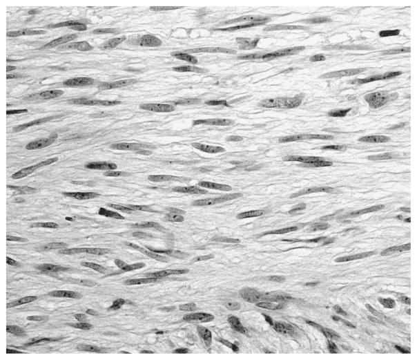
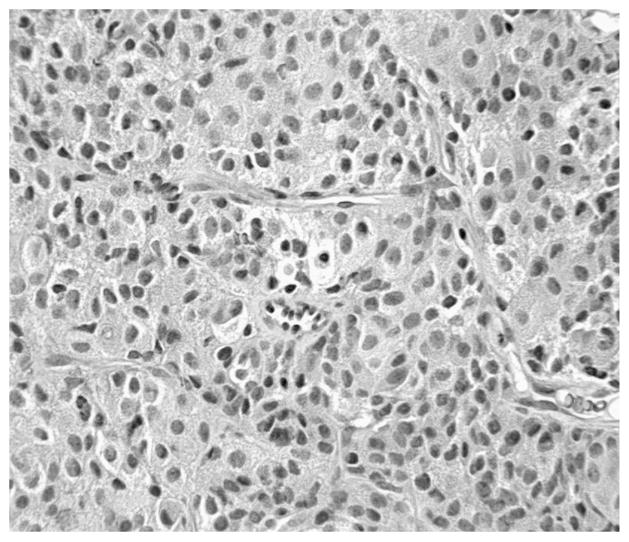
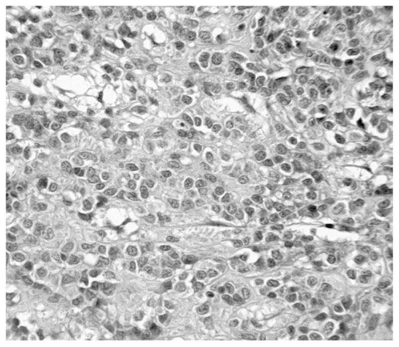
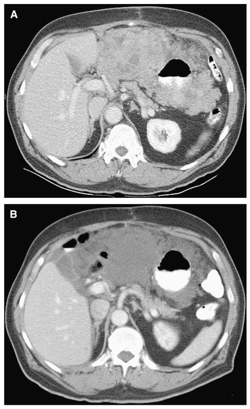
The standard of care for managing patients with gastrointestinal stromal tumors (GISTs) rapidly changed after the introduction of effective molecularly targeted therapies involving tyrosine kinase inhibitors (TKIs), such as imatinib mesylate and sunitinib malate. A better understanding of the molecular characteristics of GISTs have improved the diagnostic accuracy and led to the discovery of novel immunomarkers and new mechanisms of resistance to TKI therapy, which in turn have resulted in the development of novel treatment strategies. To address these issues, the NCCN organized a task force consisting of a multidisciplinary panel of experts in the fields of medical oncology, surgical oncology, molecular diagnostics, and pathology to discuss the recent advances, identify areas of future research, and recommend an optimal approach to care for patients with GIST at all stages of disease. The task force met for the first time in October 2003 and again in December 2006 and October 2009. This supplement describes the recent developments in the field of GIST as discussed at the October 2009 meeting.


Conflict of interest statement

The NCCN guidelines staff have no conflicts to disclose.

Figures

Figure 1
Figure 1
Spindle cell gastrointestinal stromal tumor (GIST). Typical morphology of a low-risk GIST comprised predominantly of spindle cells. This tumor was strongly KIT-positive and harbored a mutation in KIT exon 11 (H&E stain; original magnification, 400×). Courtesy of Christopher L. Corless, MD, PhD, Oregon Health & Science University.
Figure 2
Figure 2
Intermediate-risk gastrointestinal stromal tumor (GIST) comprised predominantly of epithelioid cells. The tumor was KIT-positive and contained a mutation in KIT exon 9 (H&E stain; original magnification, 400×). Courtesy of Christopher L. Corless, MD, PhD, Oregon Health & Science University.
Figure 3
Figure 3
Platelet-derived growth factor receptor alpha (PDGFRA)–mutant gastrointestinal stromal tumor (GIST). This malignant, epithelioid GIST was KIT-negative and had a mutation in PDGFRA exon 18 (H&E stain; original magnification, 400×). Courtesy of Christopher L. Corless, MD, PhD, Oregon Health & Science University.
Figure 4
Figure 4
Nomogram for predicting probabilities of 2- and 5-year recurrence-free survival. Points are assigned for size, mitotic index, and site of origin by drawing a line upward from the corresponding values to the “Points” line. The sum of these 3 points, plotted on the “Total Points” line, corresponds to predictions of 2- and 5-year recurrence-free survival. Abbreviations: HPF, high-power field; RFS, recurrence-free survival. Data from Gold JS, Gonen M, Gutierrez A, et al. Development and validation of a prognostic nomogram for recurrence-free survival after complete surgical resection of localised primary gastrointestinal stromal tumour: a retrospective analysis. Lancet Oncol 2009;10:1045–1052.
Figure 5
Figure 5
Approach for the management of very small gastric gastrointestinal stromal tumors (GISTs).* *Possible high-risk endoscopic ultrasound (EUS) features include irregular border, cystic spaces, ulceration, echogenic foci, and heterogeneity. EUS should only be considered after a thorough discussion with the patient regarding the risks and benefits. Adapted from Sepe PS, Brugge WR. A guide for the diagnosis and management of gastrointestinal stromal cell tumors. Nat Rev Gastroenterol Hepatol 2009;6:363–371. The panel included this approach as a category 2B recommendation.
Figure 6
Figure 6
A spurious “new” lesion on follow-up CT in a 41-year-old man with primary gastrointestinal stromal tumor in the small bowel who received imatinib treatment. (A, B) On pretreatment CT, a metastatic lesion (arrow) in the liver could only be detected on an unenhanced image (A) but not on the enhanced portal-venous phase image (B), because the lesion was enhanced to the same degree as the surrounding parenchyma. (C) A portal-venous phase image of CT obtained 8 weeks after treatment showed that the lesion (arrow) became clearly visible, which should not be misinterpreted as a new lesion. Courtesy of Haesun Choi, MD, The University of Texas M. D. Anderson Cancer Center.
Figure 7
Figure 7
Typical appearance of gastrointestinal stromal tumor (GIST) in a 70-year-old man with an unresectable GIST of the stomach. (A) A pretreatment CT image showed a very large hyperdense mass completely surrounding the stomach. Endoscopic biopsy was negative for malignancy. (B) The mass became hypodense and homogenous on CT obtained 8 weeks after imatinib treatment. Courtesy of Haesun Choi, MD, The University of Texas M. D. Anderson Cancer Center.
Figure 8
Figure 8
Good response to imatinib treatment in a 50-year-old man with metastatic gastrointestinal stromal tumor of the stomach. (A) A late arterial-phase image of pretreatment CT showed multiple hypervascular metastases in the liver (large arrows). Notice small tumor vessels within the mass (small arrow). (B) On CT obtained 8 weeks after treatment, the masses became hypodense (arrows) and the tumor vessels and enhancing nodules are no longer seen. Courtesy of Haesun Choi, MD, The University of Texas M. D. Anderson Cancer Center.
Figure 9
Figure 9
Increasing tumor size and spurious progression of disease in a 41-year-old man with a primary gastrointestinal stromal tumor of the small bowel who experienced a good response to imatinib treatment. (A) A portal-venous phase image of pretreatment CT showed multiple, small hyperdense metastases in the liver (arrows). (B) At 8 weeks after treatment, the lesions became homogenous and hypodense (indicating good response) but increased in size significantly (arrows). (C) At 16 weeks after treatment, the lesion in the medial segment of the left lobe decreased significantly (large arrow). Notice the lesion in the right lobe (small arrow) had continuously increased but remained hypodense. This lesion became smaller on follow-up CTs (not shown). Courtesy of Haesun Choi, MD, The University of Texas M. D. Anderson Cancer Center.
Figure 10
Figure 10
Intratumoral recurrence after imatinib in a 72-year-old man with a primary gastrointestinal stromal tumor in the duodenum. (A) An enhanced CT image obtained 12 months after treatment showed multiple, treated, hypodense metastasis in both lobes of the liver (arrow). (B, C) On follow-up CTs, a continuously increasing intratumoral nodule (arrow) was noted at 17 months (B) and 22 months (C) after treatment. Courtesy of Haesun Choi, MD, The University of Texas M. D. Anderson Cancer Center.

Similar articles

Cited by

References

    1. Nishida T, Hirota S. Biological and clinical review of stromal tumors in the gastrointestinal tract. Histol Histopathol. 2000;15:1293–1301. - PubMed
    1. Thomas RM, Sobin LH. Gastrointestinal cancer. Cancer. 1995;75:154–170. - PubMed
    1. Tran T, Davila JA, El-Serag HB. The epidemiology of malignant gastrointestinal stromal tumors: an analysis of 1,458 cases from 1992 to 2000. Am J Gastroenterol. 2005;100:162–168. - PubMed
    1. Tryggvason G, Gislason HG, Magnusson MK, et al. Gastrointestinal stromal tumors in Iceland, 1990–2003: the Icelandic GIST study, a population-based incidence and pathologic risk stratification study. Int J Cancer. 2005;117:289–293. - PubMed
    1. Goettsch WG, Bos SD, Breekveldt-Postma N, et al. Incidence of gastrointestinal stromal tumors is underestimated: results of a nation-wide study. Eur J Cancer. 2005;41:2868–2872. - PubMed

Publication types

MeSH terms