

Functional connectivity density mapping
- PMID: 20457896
- PMCID: PMC2906909
- DOI: 10.1073/pnas.1001414107
Functional connectivity density mapping
Abstract
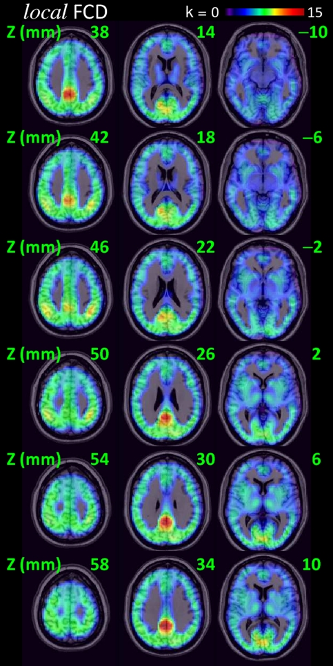
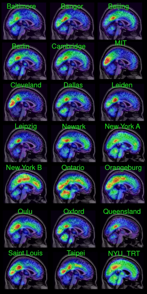
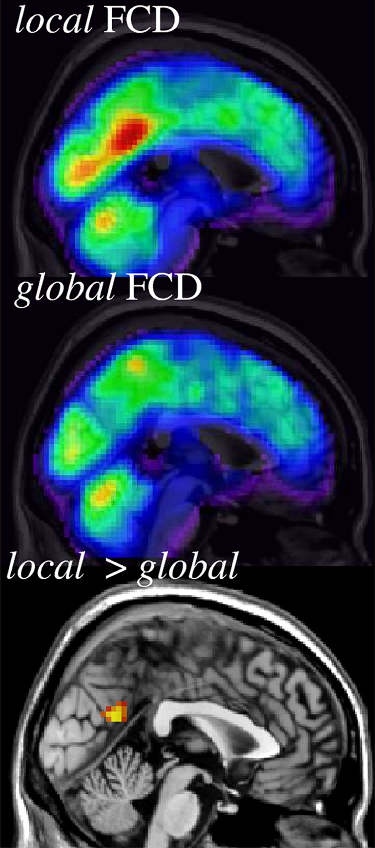
Brain networks with energy-efficient hubs might support the high cognitive performance of humans and a better understanding of their organization is likely of relevance for studying not only brain development and plasticity but also neuropsychiatric disorders. However, the distribution of hubs in the human brain is largely unknown due to the high computational demands of comprehensive analytical methods. Here we propose a 10(3) times faster method to map the distribution of the local functional connectivity density (lFCD) in the human brain. The robustness of this method was tested in 979 subjects from a large repository of MRI time series collected in resting conditions. Consistently across research sites, a region located in the posterior cingulate/ventral precuneus (BA 23/31) was the area with the highest lFCD, which suggest that this is the most prominent functional hub in the brain. In addition, regions located in the inferior parietal cortex (BA 18) and cuneus (BA 18) had high lFCD. The variability of this pattern across subjects was <36% and within subjects was 12%. The power scaling of the lFCD was consistent across research centers, suggesting that that brain networks have a "scale-free" organization.
Conflict of interest statement
The authors declare no conflict of interest.
Figures
Comment in
-
Human functional connectivity: new tools, unresolved questions.Proc Natl Acad Sci U S A. 2010 Jun 15;107(24):10769-70. doi: 10.1073/pnas.1005987107. Epub 2010 Jun 14. Proc Natl Acad Sci U S A. 2010. PMID: 20547869 Free PMC article. No abstract available.
References
-
- Watts DJ, Strogatz SH. Collective dynamics of ‘small-world’ networks. Nature. 1998;393:440–442. - PubMed
-
- Bassett DS, Bullmore E. Small-world brain networks. Neuroscientist. 2006;12:512–523. - PubMed
-
- Salvador R, et al. Neurophysiological architecture of functional magnetic resonance images of human brain. Cereb Cortex. 2005;15:1332–1342. - PubMed
Publication types
MeSH terms
Grants and funding
LinkOut - more resources
Full Text Sources
Molecular Biology Databases
