. 2010 May 25;107(21):9665-70.
doi: 10.1073/pnas.0914078107. Epub 2010 May 10.

Circadian-independent cell mitosis in immortalized fibroblasts

Affiliations

Circadian-independent cell mitosis in immortalized fibroblasts

Mijung Yeom et al. Proc Natl Acad Sci U S A. .

Abstract

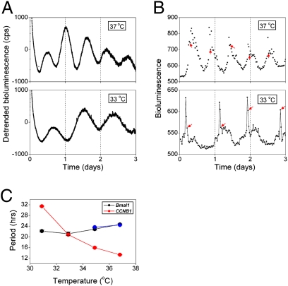
Two prominent timekeeping systems, the cell cycle, which controls cell division, and the circadian system, which controls 24-h rhythms of physiology and behavior, are found in nearly all living organisms. A distinct feature of circadian rhythms is that they are temperature-compensated such that the period of the rhythm remains constant (approximately 24 h) at different ambient temperatures. Even though the speed of cell division, or growth rate, is highly temperature-dependent, the cell-mitosis rhythm is temperature-compensated. Twenty-four-hour fluctuations in cell division have also been observed in numerous species, suggesting that the circadian system is regulating the timing of cell division. We tested whether the cell-cycle rhythm was coupled to the circadian system in immortalized rat-1 fibroblasts by monitoring cell-cycle gene promoter-driven luciferase activity. We found that there was no consistent phase relationship between the circadian and cell cycles, and that the cell-cycle rhythm was not temperature-compensated in rat-1 fibroblasts. These data suggest that the circadian system does not regulate the cell-mitosis rhythm in rat-1 fibroblasts. These findings are inconsistent with numerous studies that suggest that cell mitosis is regulated by the circadian system in mammalian tissues in vivo. To account for this discrepancy, we propose two possibilities: (i) There is no direct coupling between the circadian rhythm and cell cycle but the timing of cell mitosis is synchronized with the rhythmic host environment, or (ii) coupling between the circadian rhythm and cell cycle exists in normal cells but it is disconnected in immortalized cells.


Conflict of interest statement

The authors declare no conflict of interest.

Figures

Fig. 1.
Fig. 1.
Rhythmic CCNB1-dGluc expression in rat-1 fibroblasts is synchronized by changing the media. Rat-1 fibroblasts (0.5% confluency) stably expressing CCNB1-dGluc were subcultured in growth media and maintained in a CO2 incubator. Two days later, the growth media were replaced with recording media and luminescence was recorded. (A) Raw CCNB1-dGluc bioluminescence (counts/s) was measured for 3 days. (B) Detrended CCNB1-dGluc bioluminescence (counts/s) was obtained by subtracting the 14-h moving average from the raw data. A representative example is shown in A and B (from a total of eight dishes). (C) Two days after subculture, the growth media were replaced with recording media in one set of dishes at 0 h (black trace; n = 4) and 6 h later in another set of dishes (red trace; n = 4). Detrended (subtracted 14-h moving average) CCNB1-dGluc bioluminescence is shown. Arrows indicate the time of media change (luminescence recording start time).
Fig. 2.
Fig. 2.
Cell division occurs at the peak of the CCNB1-dGluc rhythm in rat-1 fibroblasts. Images (29-min exposures) of CCNB1-dGluc bioluminescence from rat-1 fibroblasts after a media change were collected every 30 min. Eight individual cells from two independent dishes were analyzed. (A) The relative intensity of CCNB1-dGluc expression (22 h are shown) is indicated by the color gradient, where white is high expression and black is no expression. Arrowheads indicate cells that originated from the same parent cell. (B) The optical density of CCNB1-dGluc bioluminescence in a single cell is plotted as a function of time. The times when the cell divided (one cell visibly splitting into two cells) are indicated by red arrows. (C) The phase and period of CCNB1-dGluc expression are similar in parent and daughter cells. Relative intensity of CCNB1-dGluc expression as a function of time is indicated by the color gradient, where red is high expression and green is low expression. Branch points represent the time when the parent cell split into two daughter cells.
Fig. 3.
Fig. 3.
Rhythmic Bmal1-dGluc expression in rat-1 fibroblasts is synchronized by changing the media. Rat-1 fibroblasts (0.5% confluency) stably expressing Bmal1-dGluc were subcultured in growth media and maintained in a CO2 incubator. Two days later, the growth media were replaced with recording media and luminescence was recorded. (A) Raw Bmal1-dGluc bioluminescence (counts/s) was measured for 5 days. (B) Detrended Bmal1-dGluc bioluminescence (counts/s) was obtained by subtracting the 24-h moving average from the raw data. A representative example is shown in A and B (from a total of seven dishes). (C) Two days after subculture, the growth media were replaced with recording media in one set of dishes at 0 h (black trace; n = 3) and 6 h later in another set of dishes (red trace; n = 4). Detrended (subtracted 24-h moving average) Bmal1-dGluc bioluminescence is shown. Arrows indicate the time of media change (luminescence recording start time).
Fig. 4.
Fig. 4.
Circadian and cell-cycle gene expression rhythms in rat-1 fibroblasts are independent from each other. Rat-1 fibroblasts stably transfected with Bmal1-dGluc or CCNB1-dGluc (0.5% confluency) were subcultured in growth media and maintained in a CO2 incubator. Two days later, the growth media were replaced with recording media and luminescence was recorded. Raw (A) and detrended (B) data are shown. To compare the phase of the rhythms to each other, the Bmal1-dGluc (black trace) and CCNB1-dGluc (blue trace) were recorded at the same time under the same conditions and both were detrended by subtracting the 24-h moving average. The first peak of the CCNB1-dGluc rhythm was masked by subtracting the 24-h moving average. An independent experiment showed nearly identical results.
Fig. 5.
Fig. 5.
The period of rhythmic CCNB1-dGluc expression is not temperature-compensated in rat-1 fibroblasts. Rat-1 fibroblasts, stably transfected with CCNB1-dGluc or Bmal1-dGluc, were subcultured (0.5% confluency) in growth media and maintained in a CO2 incubator. Two days later, the growth media were replaced with recording media and cells were placed in a non-CO2 incubator at 31 °C, 33 °C, 35 °C, or 37 °C. (A) Representative traces of detrended CCNB1-dGluc expression at 37 °C (14-h moving average) and 33 °C (24-h moving average) measured in LumiCycle software. (B) The optical density of CCNB1-dGluc bioluminescence in a single cell imaged at 37 °C or 33 °C. The timing of cell mitosis is indicated by red arrows. (C) The periods (hours) of the CCNB1-dGluc (red) and Bmal1-dGluc rhythms are plotted as a function of temperature (°C). Because the period of the CCNB1-dGluc rhythm varied with temperature, the following moving averages were subtracted from the raw data: 31 °C: 35 h; 33 °C: 24 h; 35 °C: 17 h; 37 °C: 14 h (n = 7 at each temperature). Raw Bmal1-dGluc bioluminescence was detrended by subtracting the 24-h moving average. We could only consistently detect rhythmic Bmal1-dGluc induced by a medium change at 35 °C and 37 °C (blue; n = 3 at each temperature). To measure the period of the circadian rhythm at low temperatures, we stimulated rat-1 fibroblasts expressing Bmal1-dGluc with dexamethasone for 3 h and then recorded bioluminescence at 31 °C, 33 °C, 35 °C, or 37 °C (n = 4 at each temperature). The periods of the dexamethasone-stimulated Bmal1-dGluc rhythms (black) were similar to the rhythms measured without dexamethasone stimulation (blue) at 35 °C and 37 °C. Data are presented as the mean ± SEM. Error bars are plotted but are smaller than the symbols. Note: At 37 °C, there was no significant difference (P = 0.61) between the periods of the CCNB1-dGluc rhythm after dexamethasone stimulation (13.03 ± 0.41 h) and after the media change (13.20 ± 0.08 h).

Similar articles

Cited by

References

    1. Pittendrigh CS. Temporal organization: Reflections of a Darwinian clock-watcher. Annu Rev Physiol. 1993;55:16–54. - PubMed
    1. Edmunds LN. Cellular and Molecular Bases of Biological Clocks: Models and Mechanisms for Circadian Timekeeping. New York: Springer; 1988.
    1. Bjarnason GA, Jordan RC, Sothern RB. Circadian variation in the expression of cell-cycle proteins in human oral epithelium. Am J Pathol. 1999;154:613–622. - PMC - PubMed
    1. Brown WR. A review and mathematical analysis of circadian rhythms in cell proliferation in mouse, rat, and human epidermis. J Invest Dermatol. 1991;97:273–280. - PubMed
    1. Smaaland R. Circadian rhythm of cell division. Prog Cell Cycle Res. 1996;2:241–266. - PubMed

Publication types

LinkOut - more resources