. 2010 May 25;107(21):9777-82.
doi: 10.1073/pnas.0914127107. Epub 2010 May 10.

Essential role of the Wnt pathway effector Tcf-1 for the establishment of functional CD8 T cell memory

Affiliations

Essential role of the Wnt pathway effector Tcf-1 for the establishment of functional CD8 T cell memory

Grégoire Jeannet et al. Proc Natl Acad Sci U S A. .

Abstract

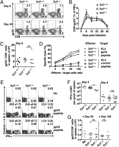
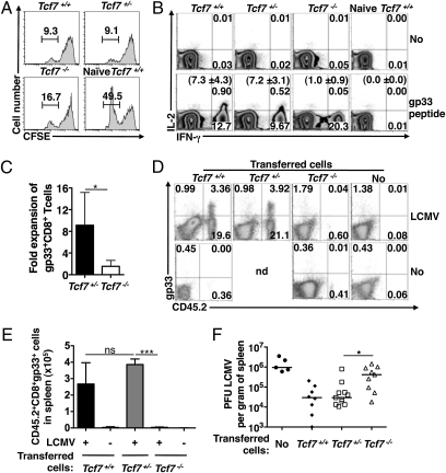
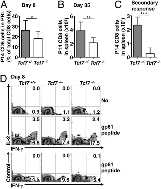
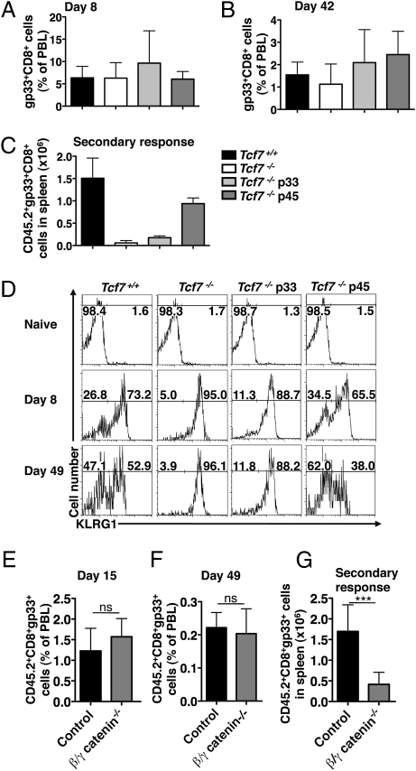
Immune protection from intracellular pathogens depends on the generation of terminally differentiated effector and of multipotent memory precursor CD8 T cells, which rapidly regenerate effector and memory cells during recurrent infection. The identification of factors and pathways involved in CD8 T cell differentiation is of obvious importance to improve vaccination strategies. Here, we show that mice lacking T cell factor 1 (Tcf-1), a nuclear effector of the canonical Wingless/Integration 1 (Wnt) signaling pathway, mount normal effector and effector memory CD8 T cell responses to infection with lymphocytic choriomeningitis virus (LCMV). However, Tcf-1-deficient CD8 T cells are selectively impaired in their ability to expand upon secondary challenge and to protect from recurrent virus infection. Tcf-1-deficient mice essentially lack CD8 memory precursor T cells, which is evident already at the peak of the primary response, suggesting that Tcf-1 programs CD8 memory cell fate. The function of Tcf-1 to establish CD8 T cell memory is dependent on the catenin-binding domain in Tcf-1 and requires the Tcf-1 coactivators and Wnt signaling intermediates beta-catenin and gamma-catenin. These findings demonstrate that the canonical Wnt signaling pathway plays an essential role for CD8 central memory T cell differentiation under physiological conditions in vivo. They raise the possibility that modulation of Wnt signaling may be exploited to improve the generation of CD8 memory T cells during vaccination or for therapies designed to promote sustained cytotoxic CD8 T cell responses against tumors.


Conflict of interest statement

The authors declare no conflict of interest.

Figures

Fig. 1.
Fig. 1.
Primary CD8 T cell response of Tcf7−/− mice to acute LCMV Infection. Tcf7+/+ (B6), Tcf7+/− and Tcf7−/− mice were infected with LCMV. (A) The antigen-specific CD8 T cell response was determined at day 8 and day 43 post-LCMV infection. Numbers indicate the percentage of peripheral blood lymphocytes (PBL) in the respective quadrant. (B) The line graph depicts the mean percentage (±SD) of gp33+ CD8+ cells among PBL at the indicated time points post-LCMV infection. (C) The absolute number of gp33+ CD8+ cells per spleen was determined at day 8 after LCMV infection. Symbols depict individual mice. (D) EL4 cells (H-2b) were pulsed with gp33 or Smcy (control) peptide and used as target cells in standard cytolysis assays using day 8 LCMV immune splenocytes as effectors. (E) Splenocytes from day-8 LCMV immune mice were stimulated in vitro with gp33 peptide before intracellular staining using IFN-γ and IL-2 or control mAbs. Density plots show gated CD8 T cells. Numbers indicate the percentage of cells in the respective quadrants. Numbers in brackets indicate the percentage (± SD or individual determinations) of IL-2+ cells among IFN-γ+ CD8+ cells from multiple determinations. (F). Recipient spleens were analyzed for infectious LCMV particles (pfus) at day 5 and day 8 postinfection. Symbols depict pfus in individual mice. (G) The number of gp33-specific CD8 cells per spleen was determined at the indicated time points post-LCMV infection. Symbols depict individual mice. (*) depict significant differences (P < 0.05); ns, not significantly different (P > 0.05) based on Student's t test.
Fig. 2.
Fig. 2.
Secondary CD8 T cell response of Tcf7−/− mice to LCMV infection. (A) B6 splenocytes were labeled with a high and with low concentration of CFSE and the latter were pulsed with gp33 peptide. A 1:1 mixture of these cells was injected into LCMV immune Tcf7+/+ (B6), Tcf7+/−, or Tcf7−/− mice around day 50 post-LCMV infection. Numbers indicate the percentage of residual CFSElo (gp33 peptide pulsed) cells relative to CFSEhi control cells in recipient spleens 3 hr after transfer. (B) Splenocytes from LCMV immune mice (around day 80) were stimulated in vitro with gp33 peptide. Density plots show gated CD8 T cells stained for intracellular IFN-γ and IL-2. Numbers indicate the percentage of cells in the respective quadrants. Numbers in brackets depict the mean percentage (± SD) of IL-2+ cells among IFN-γ+ CD8+ cells of multiple determinations. (C) The frequency of gp33-specific CD8 T cells among PBL of LCMV immune mice (>day 60 to day 70) was estimated (Tcf7+/+ 0.93 ± 0.44; Tcf7−/− 0.90 ± 0.36). These mice were challenged with gp33 peptide coupled to virus like particles and the frequency of antigen-specific CD8 T cells was determined 8 days later (Tcf7+/+ 6.66 ± 2.42, n = 5; Tcf7−/− 1.18 ± 0.67, n = 7). The bar graph depicts the mean fold expansion (± SD) of gp33-specific CD8 T cells from individual mice. (D) Splenocytes from LCMV immune Tcf7+/+, Tcf7+/−, or Tcf7−/− mice (CD45.2) (around day 85) containing 3 × 10⁴ gp33+ CD8+ cells were transferred into naïve recipients (CD45.1) followed or not (No) by LCMV infection 1 day later. Density plots show gated CD8 T cells stained with gp33 tetramer versus anti-CD45.2 at day 5 postinfection. Numbers indicate the percentage of cells in the respective quadrants. (E) The bar graph depicts the mean number (± SD) of donor-derived (CD45.2) gp33+ CD8+ T cells in recipient spleens at day 5 after infection (+) or not (−) with LCMV. (F) Naive wild-type mice received or not (No) LCMV immune cells followed by challenge infection as in D. Five days later, recipient spleens were analyzed for the abundance of infectious LCMV particles (pfu). Symbols depict individual mice. (*) and (***) depict significant differences (P < 0.05) and (P < 0.001), respectively; ns, not significantly different (P > 0.05) based on Student's t test.
Fig. 3.
Fig. 3.
Cell intrinsic role of Tcf-1 for the recall response of CD8 T cells. (A) Purified Tcf7+/− or Tcf7−/− P14 CD8 T cells (3 × 10⁴) (CD45.2) were transferred into wild-type (CD45.1/2) recipients followed by LCMV infection 1 day later. The bar graph shows the mean percentage (± SD) of P14 CD8 T cells (CD45.2) among recipient PBL at day 8 postinfection. (B) The bar graph shows the mean number (± SD) of P14 CD8 T cells in recipient spleens at day 35 to day 60 postinfection. (C) Splenocytes from primary recipients containing 3 × 10⁴ immune P14 CD8 T cells (day 42 to day 60) were transferred into secondary wild-type (CD45.1/2) recipients 1 day before LCMV challenge infection. The bar graph shows the mean number (± SD) of P14 CD8 T cells (CD45.2) in recipient spleens 5 days later. (*), (**) and (***) depict significant differences (P < 0.05), (P < 0.01) and (P < 0.001), respectively based on Student's t test. (D) Splenocytes from Tcf7+/+, Tcf7+/−, and Tcf7−/− LCMV immune mice at day 8 postinfection were stimulated in vitro with gp61 peptide. Density plots show gated CD4 T cells stained intracellularly for IFN-γ and IL-2 or using a control Ab. Numbers indicate the percentage of cells in the respective quadrants.
Fig. 4.
Fig. 4.
The catenin-binding domain in Tcf-1 and β−/γ-catenin are essential for the formation of functional CD8 T cell memory. Tcf7+/+ (B6), Tcf7−/−, Tcf7−/− p33 and Tcf7−/− p45 Tg mice were infected with LCMV. Bar graphs depict the mean percentage (± SD) of gp33+ CD8+ T cells in peripheral blood at day 8 (A) and at day 42 (B) post-LCMV infection. (C) Splenocytes from day 50 LCMV immune mice (CD45.2), containing 3 × 10⁴ gp33+ CD8+ cells, were transferred into naïve recipients (CD45.1) 1 day before LCMV infection. The bar graph depicts the mean number (± SD) of CD45.2+ gp33-specific CD8 T cells in recipient spleens 5 days later. (D) Histograms show KLRG1 expression among CD8 T cells of naïve mice and among gated gp33+ CD8+ T cells from day 8 and day 49 LCMV immune mice. Numbers indicate the percentage of cells in the respective gate. Chimeric mice with a wild-type (control) or β/γ-catenin-deficient (β/γ-catenin−/−) hematopoietic system (>90% CD45.2+) were infected with LCMV. Bar graphs depict the mean percentage (± SD) of gp33+ CD8+ T cells among CD45.2+ PBL of the indicated chimera at day 15 (E) and day 49 (F) post-LCMV infection. (G) Splenocytes from the indicated LCMV immune chimera (day 50), containing 3 × 10⁴ CD45.2+ gp33+ CD8+ T cells, were transferred into naïve recipients (CD45.1) 1 day before LCMV challenge infection. The bar graph depicts the mean number (± SD) of CD45.2+ gp33+ CD8+ T cells in recipient spleens 5 days later. (*) and (***) depict significant differences (P < 0.05) and (P < 0.001), respectively; ns, not significantly different (P > 0.05) based on Student's t test.

Similar articles

Cited by

References

    1. Stemberger C, et al. Stem cell-like plasticity of naïve and distinct memory CD8+ T cell subsets. Semin Immunol. 2009;21:62–68. - PubMed
    1. Parish IA, Kaech SM. Diversity in CD8(+) T cell differentiation. Curr Opin Immunol. 2009;21:291–297. - PMC - PubMed
    1. Mercado R, et al. Early programming of T cell populations responding to bacterial infection. J Immunol. 2000;165:6833–6839. - PubMed
    1. Kaech SM, Ahmed R. Memory CD8+ T cell differentiation: Initial antigen encounter triggers a developmental program in naïve cells. Nat Immunol. 2001;2:415–422. - PMC - PubMed
    1. Stemberger C, et al. A single naive CD8+ T cell precursor can develop into diverse effector and memory subsets. Immunity. 2007;27:985–997. - PubMed

Publication types

MeSH terms