. 2010 Jun 15;107(24):10791-8.
doi: 10.1073/pnas.0914076107. Epub 2010 May 10.

Insulin receptor functionally enhances multistage tumor progression and conveys intrinsic resistance to IGF-1R targeted therapy

Affiliations

Insulin receptor functionally enhances multistage tumor progression and conveys intrinsic resistance to IGF-1R targeted therapy

Danielle B Ulanet et al. Proc Natl Acad Sci U S A. .

Abstract

The type 1 insulin-like growth factor receptor (IGF-1R) tyrosine kinase is an important mediator of the protumorigenic effects of IGF-I/II, and inhibitors of IGF-1R signaling are currently being tested in clinical cancer trials aiming to assess the utility of this receptor as a therapeutic target. Despite mounting evidence that the highly homologous insulin receptor (IR) can also convey protumorigenic signals, its direct role in cancer progression has not been genetically defined in vivo, and it remains unclear whether such a role for IR signaling could compromise the efficacy of selective IGF-1R targeting strategies. A transgenic mouse model of pancreatic neuroendocrine carcinogenesis engages the IGF signaling pathway, as revealed by its dependence on IGF-II and by accelerated malignant progression upon IGF-1R overexpression. Surprisingly, preclinical trials with an inhibitory monoclonal antibody to IGF-1R did not significantly impact tumor growth, prompting us to investigate the involvement of IR. The levels of IR were found to be significantly up-regulated during multistep progression from hyperplastic lesions to islet tumors. Its functional involvement was revealed by genetic disruption of the IR gene in the oncogene-expressing pancreatic beta cells, which resulted in reduced tumor burden accompanied by increased apoptosis. Notably, the IR knockout tumors now exhibited sensitivity to anti-IGF-1R therapy; similarly, high IR to IGF-1R ratios demonstrably conveyed resistance to IGF-1R inhibition in human breast cancer cells. The results predict that elevated IR signaling before and after treatment will respectively manifest intrinsic and adaptive resistance to anti-IGF-1R therapies.


Conflict of interest statement

Conflict of interest statement: D.L.L. is an employee of ImClone. However, this study does not promote the ImClone drug A12.

Figures

Fig. 1.
Fig. 1.
An IGF-1R inhibitor does not significantly impair tumor growth in RIP1-Tag2 mice. (A and B) RIP1-Tag2; Rag-KO mice were treated in a 3-week intervention trial with the A12 anti–IGF-1R mAb or control antibody from 10 to 13 weeks of age (n = 13, control antibody treated mice; n = 15, A12 treated mice). Effects of A12 on tumor burden (A) and tumor number (B; *P = 0.04). (C and D) RIP1-Tag2 mice of tumor-bearing age (13 to 14 weeks) were treated in a short-term, 4-day trial with A12 or control antibody to assess effects on tumor cell apoptosis (C) as measured by TUNEL staining (*P = 0.01) and on proliferation (D) as measured by BrdU incorporation. Results represent values calculated from 15 to 20 tumor sections from three mice per group. Mean values plus SEM indicated; two-tailed, unpaired t test (with Welch correction for C and D) for statistical significance. (E) RIP1-Tag2 mice were treated with 1 mg A12 or control antibody and analyzed 6 h after injection. Pools of seven to eight tumors from three to four mice per group were analyzed by Western blotting for protein levels of IGF-1R and phosphorylated AKT (pAKT) and MEK (pMEK); blots were stripped and reprobed with antibodies to total AKT and MEK1/2 for protein load control.
Fig. 2.
Fig. 2.
The IR isoform A, a second signaling receptor for IGF-II, is expressed throughout the stages of RIP1-Tag2 tumorigenesis. (A) RT-PCR to detect IR isoform expression in mRNA isolated from normal islets and preneoplastic islets and tumors from RIP1-Tag2 mice. IR-A is expressed in normal islets and at all stages of RIP1-Tag2 tumorigenesis. (B) Expression of IR and IGF-1R protein is up-regulated in the multistage PNET tumorigenesis pathway. Protein extracts from normal, nontransgenic islets and from hyperplastic, angiogenic islets, and tumors of RIP1-Tag2 mice were immunoblotted with antibodies to the IR and IGF-1R β chain and to β-actin as a loading control. (C) IGF-II activates the IR in β tumor cells isolated from a RIP1-Tag2 tumor. βTC4 cells were stimulated with or without IGF-II and phosphorylated IR protein visualized after immunoprecipitation of protein extracts with an antibody to the IR β chain followed by immunoblotting with an anti-pTyr antibody.
Fig. 3.
Fig. 3.
Tissue-specific knockout of IR in β cells impairs PNET tumorigenesis. (A–C) Double transgenic RIP1-Tag2, RIP-Cre mice of the indicated IR genotypes were analyzed at 13 weeks of age: β-IR WT (+/+); heterozygous β-IRKO (fl/+); and β-IRKO (fl/fl). (A) Effects on tumor burden; n = 13 (+/+), n = 22 (fl/+), n = 37 (fl/fl); *P = 0.001 compared with β-IRwt mice, *P = 0.02 compared with fl/+ mice (B and C) Effects on apoptosis (B; *P = 0.006) and proliferation (C; P = 0.33); n = 17 tumor sections from five mice (+/+), n = 18 tumor sections from eight mice (fl/fl). Bars represent mean values ± SEM. Two-tailed, unpaired t test (with Welch correction for B and C). (D) Selection against complete loss of insulin/IGF signaling receptors in tumor cells. IR and IGF-1R protein levels in pooled tumor extracts from RIP1-Tag2, β-IRKO mice (Left). Tumor pools consist of equal protein amounts from 5 tumors from 4 mice (IR +/+) and 10 tumors from 10 mice (IR fl/fl). Recombination of IR gene in different stages of islet tumor development in a RIP1-Tag2; β-IRKO mouse (Center). IR and IGF-1R protein levels in islet pools from β-IRKO mice (Right; αβ precursor depicted owing to low levels of processed, mature protein).
Fig. 4.
Fig. 4.
Loss of IR expression sensitizes PNET tumors to IGF-1R inhibition. (A) RIP1-Tag2; β-IRKO mice were treated with 1 mg A12 or control antibody and analyzed 6 h after injection. Pools of four tumors from at least two mice per group were analyzed. As a comparison, protein extracts from similarly treated WT RIP1-Tag2 mice were analyzed alongside. (B and C) RIP1-Tag2; β-IRKO mice were treated in a 2-week intervention trial with A12 or control antibody from 12 to 14 weeks of age (n = 13 for control antibody, n = 17 for A12 treated mice). Effects on tumor burden (B; *P = 0.02) and tumor number (C; *P = 0.0005). Mann-Whitney test for statistical significance. (D and E) RIP1-Tag2; β-IRKO mice were treated with a single dose of A12 antibody and effects on tumor cell apoptosis (D; P = 0.1) as measured by TUNEL staining (≥15 tumor sections from four to seven mice per group analyzed), and proliferation (E; *P = 0.02) as measured by BrdU incorporation (≥22 tumor sections from six to seven mice per group analyzed) were analyzed after 24 h. Mean values +SEM are indicated. Two-tailed, unpaired t test (with Welch correction for BrdU analysis).
Fig. 5.
Fig. 5.
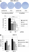
Enhanced growth inhibition of breast cancer cells by IGF-1R inhibitor in the setting of IR knockdown. (A) MDA-MB-231 cells stably transduced with an NS or IR-targeting shRNA were grown in soft agar in the presence of control or A12 antibodies. Images (Upper) and values (Lower) from two independent experiments, repeated in triplicate are depicted; Mean values plus SD are indicated and expressed as percentage colony formation relative to that of NS-shRNA expressing, control antibody treated cells; *P < 0.0001, **P < 1 × 10⁻⁵, ***P < 1 × 10⁻⁶, ****P < 1 × 10⁻¹¹; two-tailed, unpaired t test. (B) MCF-7 cells were transfected with control or IR siRNAs and serum-starved cells were stimulated 48 h later with 10 nM IGF-I or 20 nM IGF-II or left unstimulated in the presence of control or A12 antibodies (100 nM) for an additional 48 h. Cell growth was assessed by incubation with MTT and values normalized relative to growth of control treated, unstimulated cells. Mean values plus SEM are indicated. Results representative of four independent experiments performed in triplicate. *P < 0.01, **P < 0.001, two-tailed, unpaired t test. (Magnification: A, ×3.4.)

Comment in

  • An unhelpful hand.
    Atkinson S. Atkinson S. Nat Rev Cancer. 2010 Jul;10(7):453. doi: 10.1038/nrc2883. Nat Rev Cancer. 2010. PMID: 20589970 No abstract available.
  • Anticancer drugs: An unhelpful hand.
    Atkinson S. Atkinson S. Nat Rev Drug Discov. 2010 Jul;9(7):518. doi: 10.1038/nrd3211. Nat Rev Drug Discov. 2010. PMID: 20592746 No abstract available.

References

    1. Samani AA, Yakar S, LeRoith D, Brodt P. The role of the IGF system in cancer growth and metastasis: Overview and recent insights. Endocr Rev. 2007;28:20–47. - PubMed
    1. Pollak MN, Schernhammer ES, Hankinson SE. Insulin-like growth factors and neoplasia. Nat Rev Cancer. 2004;4:505–518. - PubMed
    1. Khandwala HM, McCutcheon IE, Flyvbjerg A, Friend KE. The effects of insulin-like growth factors on tumorigenesis and neoplastic growth. Endocr Rev. 2000;21:215–244. - PubMed
    1. Ullrich A, et al. Insulin-like growth factor I receptor primary structure: Comparison with insulin receptor suggests structural determinants that define functional specificity. EMBO J. 1986;5:2503–2512. - PMC - PubMed
    1. Frasca F, et al. Insulin receptor isoform A, a newly recognized, high-affinity insulin-like growth factor II receptor in fetal and cancer cells. Mol Cell Biol. 1999;19:3278–3288. - PMC - PubMed

Publication types

MeSH terms

LinkOut - more resources