. 2010 May 25;107(21):9891-6.
doi: 10.1073/pnas.0911854107. Epub 2010 May 10.

Role of dimer interface in activation and desensitization in AMPA receptors

Affiliations

Role of dimer interface in activation and desensitization in AMPA receptors

Jennifer Gonzalez et al. Proc Natl Acad Sci U S A. .

Abstract

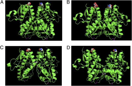
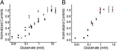
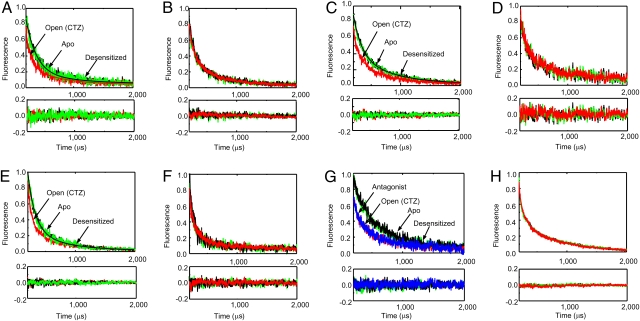
The conversion of chemical to electrical signals by the AMPA receptors is the key step by which these proteins control cognitive and motor responses. Here, we have used luminescence resonance energy transfer (LRET) to gain insight into the conformational changes induced by glutamate binding in the agonist-binding domain in functional AMPA receptors expressed in oocytes and HEK-293 cells. The LRET-based distances indicate that the interface between the upper lobes of the agonist-binding domain within a dimer is in a decoupled state in the unligated Apo state of the receptor. Agonist binding results in the formation of the dimer interface in the open-channel form of the receptor. In the continued presence of glutamate when the receptor is primarily in the desensitized state, the dimer interface is decoupled, confirming that the decoupling of the dimer interface leads to channel closure. The LRET distances also indicate that the dimer interface is preformed before activation in the L484Y mutation and also is formed in the antagonist (ZK200775)-bound form of the AMPA receptor. These results suggests that, although the preformation of the interface is not sufficient to drive channel activation, it could play a role in the energetics of activation and hence modulation of the receptor by auxiliary proteins or small molecules.


Conflict of interest statement

The authors declare no conflict of interest.

Figures

Fig. 1.
Fig. 1.
Crystal structures of AMPA receptor ligand-binding domain (GluR2-S1S2) showing the dimer interface. The structures are (A) in the Apo state, (B) in the glutamate-bound state, (C) glutamate-bound in the presence of cyclothiazide, and (D) the S729C structure thought to represent the desensitized dimer interface. Residue 740 (741 in GluR4) in GluR2-S1S2, which was tagged with the donor:acceptor fluorophore in the LRET measurements, is highlighted as spheres, and the linker region where the transmembrane segments are attached is shown in magenta. All the structures show an intact dimer interface; the only structure that is different in terms of the interface, D, is shown in the GluR2-S1S2-S729C structure.
Fig. 2.
Fig. 2.
Dose–response curve showing the dependence of the maximum current as a function of glutamate concentration. (A) ΔN-GluR4 receptors (red), ΔN*-GluR4-G740C (blue), ΔN*-GluR4-S741C (green), and ΔN*-GluR4-S742C (black) receptors expressed in oocytes with varying concentrations of glutamate in 100 μM cyclothiazide. (B) Wild-type GluR4-flip receptors (red) and ΔN*-GluR4-Th-S741C-Th receptors (black) expressed in HEK-293 cells. All currents were recorded in the presence of cyclothiazide and normalized to currents mediated by 10 mM glutamate.
Fig. 3.
Fig. 3.
The LRET lifetimes measured at 510 nm for donor:acceptor-tagged receptors. Lifetimes are shown for (A) ΔN*-GluR4-G740C, (B) ΔN*-GluR4-G740C-L484Y, (C) ΔN*-GluR4-S741C, (D) ΔN*-GluR4-S741C-L484Y, (E) ΔN*-GluR4-S742C, and (F) ΔN*-GluR4-S742C-L484Y receptors expressed in oocytes in the absence of agonists (black), with 10 mM glutamate (green), and with 10 mM glutamate in the presence of 100 μM cyclothiazide (red). Lifetimes after subtraction of the residual lifetimes upon thrombin digestion for (G) ΔN*-GluR4-Th-S741-Th and (H) ΔN*-GluR4-Th-S741-Th-L484Y receptors expressed in HEK-293 cells in the absence of agonists (black), with 10 mM glutamate (green), with 10 mM glutamate in the presence of 100 μM cyclothiazide (red), and in the presence of 200 nM antagonist ZK200775 (blue).
Fig. 4.
Fig. 4.
Proposed mechanism showing changes in the ligand-binding domain associated with the resting state, activation, and desensitization. Agonist binding to the ligand-binding domain induces cleft closure in the ligand-binding domain, pulling apart the linker to the channel segments opening the channel (activation). The open-channel form is transiently stabilized through transiently formed dimer interface interactions at the ligand-binding domain. Stress on the linker domain eventually results in decoupling of the ligand-binding domain dimer interface interactions, leading to channel closure (desensitization).

Similar articles

Cited by

References

    1. Hollmann M, Heinemann S. Cloned glutamate receptors. Annu Rev Neurosci. 1994;17:31–108. - PubMed
    1. Nakanishi S, Masu M. Molecular diversity and functions of glutamate receptors. Annu Rev Biophys Biomol Struct. 1994;23:329–348. - PubMed
    1. Madden DR. The structure and function of glutamate receptor ion channels. Nat Rev Neurosci. 2002;3:91–101. - PubMed
    1. Mayer ML. Glutamate receptor ion channels. Curr Opin Neurobiol. 2005;15:282–288. - PubMed
    1. Mayer ML. Glutamate receptors at atomic resolution. Nature. 2006;440:456–462. - PubMed

Publication types

Substances