. 2010 May 25;107(21):9861-6.
doi: 10.1073/pnas.0912877107. Epub 2010 May 10.

A molecular mechanism underlying the neural-specific defect in torsinA mutant mice

Affiliations

A molecular mechanism underlying the neural-specific defect in torsinA mutant mice

Connie E Kim et al. Proc Natl Acad Sci U S A. .

Abstract

A striking but poorly understood feature of many diseases is the unique involvement of neural tissue. One example is the CNS-specific disorder DYT1 dystonia, caused by a 3-bp deletion ("DeltaE") in the widely expressed gene TOR1A. Disease mutant knockin mice (Tor1a(DeltaE/DeltaE)) exhibit disrupted nuclear membranes selectively in neurons, mimicking the tissue specificity of the human disease and providing a model system in which to dissect the mechanisms underlying neural selectivity. Our in vivo studies demonstrate that lamina-associated polypeptide 1 (LAP1) and torsinB function with torsinA to maintain normal nuclear membrane morphology. Moreover, we show that nonneuronal cells express dramatically higher levels of torsinB and that RNAi-mediated depletion of torsinB (but not other torsin family members) causes nuclear membrane abnormalities in Tor1a(DeltaE/DeltaE) nonneuronal cells. The Tor1a(DeltaE/DeltaE) neural selective phenotype therefore arises because high levels of torsinB protect nonneuronal cells from the consequences of torsinA dysfunction, demonstrating how tissue specificity may result from differential susceptibility of cell types to insults that disrupt ubiquitous biological pathways.


Conflict of interest statement

The authors declare no conflict of interest.

Figures

Fig. 1.
Fig. 1.
LAP1 and torsinA function in a common molecular pathway in neurons. (A) LAP1 is broadly expressed in neural and nonneural murine embryonic tissue. Protein lysates from E16 wild-type tissues probed with anti-LAP1 (Upper) and anti-glyceraldehyde-3-phosphate dehydrogenase (GAPDH; Lower) antibodies. (B) LAP1 protein expression is abolished by a gene-trap cassette. Protein lysates from Tor1aip1+/+, Tor1aip1+/−, Tor1aip1−/− E18 mouse brain probed with anti-LAP1 (Upper) and anti-GAPDH (Lower) antibodies. The arrow indicates a nonspecific cross-reactive band. (C) Loss of LAP1 causes nuclear envelope abnormalities in E18 neural tissue. (Scale bar, 0.5 μm.) The nucleus (N) is marked. (D) Loss of torsinA or LAP1 causes the same ultrastructural defect of neuronal nuclear membranes. Reconstituted 3D EM tomographic images (in color) viewed from the top, side, and base of neuronal nuclear membrane abnormality (“bleb”) from Tor1a−/− and Tor1aip1−/− mice. The 3D reconstruction (in color) of the inner nuclear membrane and associated bleb is shown in the context of the EM section from which they reside. The nucleus (N) is marked.
Fig. 2.
Fig. 2.
Loss of LAP1 function causes NE blebs in nonneuronal tissues. (A) Absence of LAP1 generates blebs in E18 nonneural tissues. (Scale bar, 0.5 μm.) The nucleus (N) is marked. (B) Summary of bleb frequencies in neural and nonneural tissues in E18 LAP1 null embryos. Error bars = SEM of bleb frequencies from three independent experiments (n = 3) performed on tissues derived from three independent sets of mice. (C) Elimination of torsinA function in Tor1aip1+/− nonneuronal cells (MEFs) leads to bleb formation. (Scale bar, 0.5 μm.) The nucleus (N) is marked.
Fig. 3.
Fig. 3.
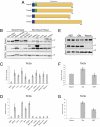
Torsin family members exhibit distinct expression patterns in neuronal and nonneuronal cells. (A) Cartoon diagram illustrating the torsin protein family. The key features of torsinA, torsinB, torsin2, and torsin3 are shown, including the signal sequence (SS; blue), hydrophobic domain (HP; green), and AAA domain (yellow). The number of amino acids is indicated for each protein. (B) The expression patterns of torsinA and torsinB proteins are opposite in neural and nonneural whole tissue. Levels of torsinB protein are clearly higher in nonneural tissue, whereas levels of torsinA protein are generally higher in neural tissue. Whole tissue protein lysates from E16 wild-type embryos probed with anti-torsinA (Top), anti-torsinB (Middle; 34 kDa), and anti-GAPDH (Bottom) antibodies. The E16 time point was chosen because this is the age when nuclear membrane blebs begin to form. (C) Levels of Tor2a mRNA are similar in neural and nonneural tissue. Quantitative RT-PCR was performed using RNA derived from E16 wild-type embryos. Ct value of Tor2a in each tissue was normalized to the Ct value of GAPDH. Relative fold change was obtained by comparing the normalized Ct value for a designated tissue to the one for heart. Error bars = SEM of fold change from three independent experiments (n = 3) performed on tissue derived from three independent sets of mice. (D) Levels of Tor3a mRNA are markedly higher in nonneural tissue than in neural tissue. Quantitative RT-PCR was performed as in C. (E) Neurons express markedly less torsinB protein than nonneuronal cell types. Whole cell protein lysates from primary cultures of wild-type MEFs, glia, or cortical neurons probed with anti-torisnA (Top), anti-torsinB (Middle), and anti-histone 3 (Bottom, loading control) antibodies. (F) Neurons and nonneuronal cell types express similar levels of Tor2a mRNA. Quantitative RT-PCR was performed using RNA extracted from primary cultures of wild-type MEFs, glia, or cortical neurons. Ct value of Tor2a for each cell type was normalized to the Ct value of β-actin. Relative fold change was obtained by comparing the normalized Ct value for a designated cell type to the one for neurons. Error bars = SEM of fold change from three independent experiments (n = 3) performed on cells derived from three independent sets of cultures. (G) Neurons express markedly less Tor3a mRNA than nonneuronal cell types. Quantitative RT-PCR was performed as in F.
Fig. 4.
Fig. 4.
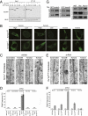
TorsinB maintains normal nuclear membrane morphology in torsinA knockout and ΔE-knockin nonneuronal cells. (A) All four torsin family members coimmunoprecipitate (co-IP) with LAP1. CAD cells were cotransfected with V5-LAP1 and each of GFP-tagged torsin family members. LAP1 was immunoprecipitated from whole cell lysates of these transfections with anti-V5 antibody, and the immunoprecipitation (IP) was separated using SDS/PAGE and probed with anti-GFP and anti-V5 antibodies. TA, torsinA; TB, torsinB; T2, torsin2; T3, torsin3; ER, GFP with ER signal sequence and KDEL retention signal. Note that ER-localized GFP (ER) does not co-IP with LAP1, and GFP-torsin does not co-IP in the absence of LAP1 (only torsin2 is shown here). (B) Loss of LAP1 selectively alters the subcellular localization of substrate trap forms of torsinA and torsinB. MEF cells derived from wild-type (Top) or Tor1aip1−/− (Lower) embryos were electroporated with GFP-tagged substrate trapping mutant torsins. Substrate trap forms of torsinA and torsinB exhibit nuclear membrane accumulation that requires LAP1. This is particularly notable for cells expressing low levels (“low”) of torsinA or torsinB. The subcellular localization of substrate trap forms of torsin2 or torsin3 was not altered in Tor1aip1−/− cells. (C) Selective formation of NE blebs following the knockdown of torsinB in Tor1a−/− MEFs. Tor1a/− and wild-type MEFs were transduced with lentivirus expressing shRNA to knock down torsinB, torsin2, torsin3, or a control shRNA and assessed by EM. Knockdown of torsinB caused NE blebs only in Tor1a/− cells. Knockdown of torsin2 or torsin3 did not cause NE blebs. The nucleus (N) is marked. (Scale bar, 0.5 μm.) (D) Selective formation of NE blebs following the knockdown of torsinB in Tor1a−/− and Tor1aΔE/ΔE MEFs. Quantification of data from experiments shown in Fig. 4 C and E. Error bars = SEM of bleb frequencies from three independent experiments (n = 3). (E) Selective formation of NE blebs following the knockdown of torsinB in Tor1aΔE/ΔE MEFs. Experiment was performed as in C. The nucleus (N) is marked. (Scale bar, 0.5 μm.) (F) Selective enhancement of NE bleb formation following the knockdown of torsinB in Tor1aΔE/ΔE neurons. Graphical illustration of data described in main text. Error bars = SEM of bleb frequencies from three independent experiments (n = 3). (G) The relative amounts of torsinA and torsinB are strikingly different in neuronal and nonneuronal cell types. Anti-torsinA and anti-torsinB antibodies were normalized using cell lysates transfected with either GFP-torsinA or GFP-torsinB (Left). These normalized conditions were used to assess the relative amounts of torsinA and torsinB in cell lysates derived from primary cultures of wild-type MEFs, glia, or neurons (Right).

Similar articles

Cited by

References

    1. Kustedjo K, Bracey MH, Cravatt BF. TorsinA and its torsion dystonia-associated mutant forms are lumenal glycoproteins that exhibit distinct subcellular localizations. J Biol Chem. 2000;275:27933–27939. - PubMed
    1. Naismith TV, Heuser JE, Breakefield XO, Hanson PI. TorsinA in the nuclear envelope. Proc Natl Acad Sci USA. 2004;101:7612–7617. - PMC - PubMed
    1. Ozelius LJ, et al. The early-onset torsion dystonia gene (DYT1) encodes an ATP-binding protein. Nat Genet. 1997;17:40–48. - PubMed
    1. Giles LM, Li L, Chin LS. Printor, a novel torsinA-interacting protein implicated in dystonia pathogenesis. J Biol Chem. 2009;284:21765–21775. - PMC - PubMed
    1. Goodchild RE, Dauer WT. The AAA+ protein torsinA interacts with a conserved domain present in LAP1 and a novel ER protein. J Cell Biol. 2005;168:855–862. - PMC - PubMed

Publication types