. 2010 May 25;107(21):9879-84.
doi: 10.1073/pnas.1004676107. Epub 2010 May 10.

Loss of leucine-rich repeat kinase 2 causes impairment of protein degradation pathways, accumulation of alpha-synuclein, and apoptotic cell death in aged mice

Affiliations

Loss of leucine-rich repeat kinase 2 causes impairment of protein degradation pathways, accumulation of alpha-synuclein, and apoptotic cell death in aged mice

Youren Tong et al. Proc Natl Acad Sci U S A. .

Abstract

Mutations in leucine-rich repeat kinase 2 (LRRK2) are the most common genetic cause of Parkinson's disease. LRRK2 is a large protein containing a small GTPase domain and a kinase domain, but its physiological role is unknown. To identify the normal function of LRRK2 in vivo, we generated two independent lines of germ-line deletion mice. The dopaminergic system of LRRK2(-/-) mice appears normal, and numbers of dopaminergic neurons and levels of striatal dopamine are unchanged. However, LRRK2(-/-) kidneys, which suffer the greatest loss of LRRK compared with other organs, develop striking accumulation and aggregation of alpha-synuclein and ubiquitinated proteins at 20 months of age. The autophagy-lysosomal pathway is also impaired in the absence of LRRK2, as indicated by accumulation of lipofuscin granules as well as altered levels of LC3-II and p62. Furthermore, loss of LRRK2 dramatically increases apoptotic cell death, inflammatory responses, and oxidative damage. Collectively, our findings show that LRRK2 plays an essential and unexpected role in the regulation of protein homeostasis during aging, and suggest that LRRK2 mutations may cause Parkinson's disease and cell death via impairment of protein degradation pathways, leading to alpha-synuclein accumulation and aggregation over time.


Conflict of interest statement

The authors declare no conflict of interest.

Figures

Fig. 1.
Fig. 1.
Age-dependent renal atrophy in LRRK2−/− mice. (A) Targeting strategy for generation of LRRK2 KO1 mice. The locations of the 5′ and 3′ external probes used for Southern blotting are indicated. Restriction site: N, NheI; S, SphI. (B and C) Southern blotting of tail genomic DNAs using 5′ probe shows germ-line transmission of targeted allele (B) and KO1 allele (C). Tail genomic DNAs were digested with NheI. The 11.5-kb band represents the WT allele, whereas the 5.4-kb and 3.7-kb bands represent the targeted allele and KO1 allele, respectively. (D) Northern blotting of total RNAs from brains of KO1 mice shows absence and reduction of LRRK2 mRNAs in −/− and +/− mice, respectively. A 406-bp cDNA fragment spanning exons 1–5 of LRRK2 was used as a probe. 18S rRNAs were used as loading control. (E) Western blotting indicates absence of LRRK2 in the brain of −/− mice. Reprobing of the same membranes with an antibody specific for spectrin was used as loading control. (F) Compared with WT controls, kidneys (fresh, nonperfused) from 20-month-old LRRK2−/− mice are significantly smaller and darker and weigh ~30% less, whereas kidneys (perfused) from 10-week-old LRRK2−/− mice are similar in size and weight to WT controls. n = 8 Kidneys per genotypic group. (G) Quantitative RT-PCR showing relative expression levels of LRRK1 and LRRK2 mRNAs, after normalized to an internal control TATA-binding protein (TBP) mRNA, in the brain and kidney of WT mice (n = 3). All data are expressed as mean ± SEM. ns, Not significant. **P < 0.01.
Fig. 2.
Fig. 2.
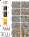
Accumulation and aggregation of α-synuclein and ubiquitinated proteins in absence of LRRK2. (A) Representative Western blots showing dramatically increased levels of α-synuclein monomers (soluble α-syn) in Triton X-100–soluble fractions and HMW α-synuclein species (HMW α-syn) and ubiquitin-positive proteins (HMW ubi+) in Triton X-100 insoluble fractions in kidneys of 20-month-old LRRK2−/− mice. Ponceau S staining is used as loading control, as levels of β-actin are altered in LRRK2−/− kidneys. (B) Quantification of Triton-soluble α-synuclein monomers and insoluble HMW α-synuclein and ubiquitin-positive proteins (bracketed region) from Western blots as shown in A. (C) Immunohistochemical analysis shows widely distributed cytosolic granules or inclusions immunoreactive to α-synuclein-, phospho–α-synuclein–specific (pS129), or ubiquitin-specific antibody in boxy cells of renal tubules in kidneys of 20-month-old LRRK2−/− mice. (All scale bars, 20 μm.) (D) Relative areas of granules immunoreactive to α-synuclein–specific (α-syn), phospho–α-synuclein–specific (pS129), or ubiquitin (ubi)–specific antibody as shown in C were estimated using the ImageJ program (National Institutes of Health). Data in all panels are expressed as mean ± SEM. *P < 0.05; **P < 0.01; ***P < 0.001.
Fig. 3.
Fig. 3.
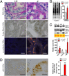
Impairment of the autophagy–lysosomal pathway and increases in oxidative damage. (A) Periodic Acid-Schiff staining of cross sections of kidneys shows thicker and darker pink staining in renal tubules and the presence of widely distributed brown granules in boxy cells of renal tubules of 20-month-old LRRK2−/− mice compared with WT controls (+/+). Nuclei were counterstained with hematoxylin (blue). Sudan Black B staining reveals, in renal tubules of LRRK2−/− mice, widely distributed dark brown granules that show bright autofluorescence (orange for merged fluorescence) in the absence of any primary and secondary antibodies. (B) Elevated levels of protein carbonyls (OxyBlot) in Triton X-100–insoluble fractions of kidneys from 20-month-old LRRK2−/− mice. Equal loading of total proteins was confirmed by Ponceau S–staining of the membranes. (C) Western blotting shows dramatically decreased levels of autophagosome marker LC3-II (some LC3-II signals are detected when more proteins were loaded and longer exposure was used) and increased levels of an autophagy substrate p62 in Triton X-100–insoluble fractions of kidneys from LRRK2−/− mice. (D) Immunohistochemical analysis revealed the presence of granular aggregates or inclusions immunoreactive to p62-specific antibody in the deeper layer of the renal cortex of LRRK2−/− mice. Relative areas of granules immunoreactive to p62 antibody were estimated using the ImageJ program (National Institutes of Health). (All scale bars, 20 μm.) Data in all panels are expressed as mean ± SEM. *P < 0.05; ***P < 0.001.
Fig. 4.
Fig. 4.
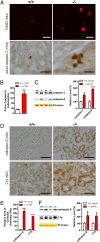
Increases in apoptotic cell death and inflammatory responses. (A) TUNEL and immunohistochemical analysis shows increased numbers of TUNEL-positive cells and densely stained active caspase-3–immunoreactive cells, respectively, in kidneys of 20-month-old LRRK2−/− mice. (B) Quantification of active caspase-3–immunoreactive cells shows dramatic increases of apoptotic cells in LRRK2−/− mice. (C) Western analysis shows increased levels of active caspase-3 and caspase-6 in Triton X-100–soluble fractions of LRRK2−/− mice. (D) Immunohistochemical analysis reveals up-regulation of cathepsin S and complement C1q, two commonly used inflammation markers, in LRRK2−/− mice. (E) Relative areas of granules immunoreactive to cathepsin S- or C1q-specific antibody were estimated using the ImageJ program (National Institutes of Health). (F) Western blotting confirms the elevated level of cathepsin S and C1q in Triton X-100–insoluble fractions of kidneys from 20-month-old LRRK2−/− mice. (Scale bars, 20 μm for ×64 and ×100, and 50 μm for ×40.) Data in all panels are expressed as mean ± SEM. *P < 0.05; **P < 0.01; ***P < 0.001.

Similar articles

Cited by

References

    1. Spillantini MG, et al. Alpha-synuclein in Lewy bodies. Nature. 1997;388:839–840. - PubMed
    1. Paisán-Ruíz C, et al. Cloning of the gene containing mutations that cause PARK8-linked Parkinson's disease. Neuron. 2004;44:595–600. - PubMed
    1. Zimprich A, et al. Mutations in LRRK2 cause autosomal-dominant parkinsonism with pleomorphic pathology. Neuron. 2004;44:601–607. - PubMed
    1. Marín I. The Parkinson disease gene LRRK2: Evolutionary and structural insights. Mol Biol Evol. 2006;23:2423–2433. - PubMed
    1. Deng J, et al. Structure of the ROC domain from the Parkinson's disease-associated leucine-rich repeat kinase 2 reveals a dimeric GTPase. Proc Natl Acad Sci USA. 2008;105:1499–1504. - PMC - PubMed

Publication types

Substances