. 2010 May 18;107(20):9060-5.
doi: 10.1073/pnas.1001569107. Epub 2010 May 10.

Archaeopteryx feathers and bone chemistry fully revealed via synchrotron imaging

Affiliations

Archaeopteryx feathers and bone chemistry fully revealed via synchrotron imaging

U Bergmann et al. Proc Natl Acad Sci U S A. .

Abstract

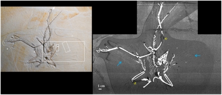
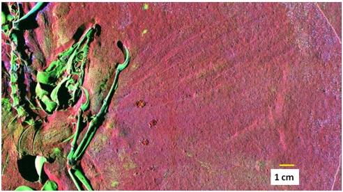
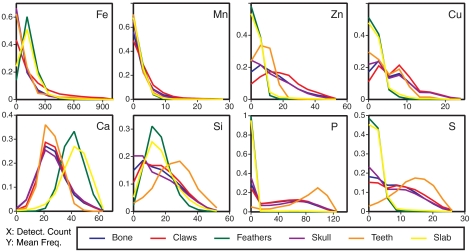
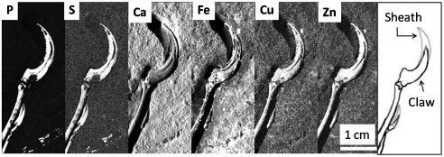
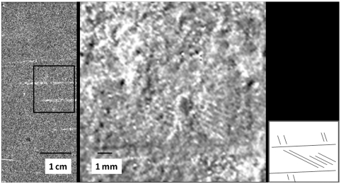
Evolution of flight in maniraptoran dinosaurs is marked by the acquisition of distinct avian characters, such as feathers, as seen in Archaeopteryx from the Solnhofen limestone. These rare fossils were pivotal in confirming the dinosauria-avian lineage. One of the key derived avian characters is the possession of feathers, details of which were remarkably preserved in the Lagerstätte environment. These structures were previously simply assumed to be impressions; however, a detailed chemical analysis has, until now, never been completed on any Archaeopteryx specimen. Here we present chemical imaging via synchrotron rapid scanning X-ray fluorescence (SRS-XRF) of the Thermopolis Archaeopteryx, which shows that portions of the feathers are not impressions but are in fact remnant body fossil structures, maintaining elemental compositions that are completely different from the embedding geological matrix. Our results indicate phosphorous and sulfur retention in soft tissue as well as trace metal (Zn and Cu) retention in bone. Other previously unknown chemical details of Archaeopteryx are also revealed in this study including: bone chemistry, taphonomy (fossilization process), and curation artifacts. SRS-XRF represents a major advancement in the study of the life chemistry and fossilization processes of Archaeopteryx and other extinct organisms because it is now practical to image the chemistry of large specimens rapidly at concentration levels of parts per million. This technique has wider application to the archaeological, forensic, and biological sciences, enabling the mapping of "unseen" compounds critical to understanding biological structures, modes of preservation, and environmental context.


Conflict of interest statement

The authors declare no conflict of interest.

Figures

Fig. 1.
Fig. 1.
SRS-XRF map of the phosphorous distribution in the Thermopolis Archaeopteryx. This map clearly shows the splay of the rachises from the flight feathers (Blue Arrows) and the reconstructed areas (Yellow Arrows). The Inset shows visible light photograph where, although striking feather impressions exist, the rachises are not discernible. The Inset also shows the location of several point analyses (White Dots) and regions highlighted for image analysis (White Rectangles).
Fig. 2.
Fig. 2.
False color SRS-XRF map of Archaeopteryx. Color code is calcium, red; Zn, green; and Mn, blue. Higher intensities correspond to higher concentrations; see Table 1 for concentrations. Host rock is limestone, and hence high calcium surrounds the fossil. Blue flecks on the surface result from the presence of tiny precipitates of Mn(IV) oxide minerals that are ubiquitous along bedding planes and within fractures. There is some zinc associated with the mineral precipitates, but almost all of the Zn inventory in this image is associated with the Archaeopteryx bone material. Zinc apparently was present in appreciable concentrations in the original bone (as in many extant organisms) and has been well sequestered within the bone over 150 million years of burial.
Fig. 3.
Fig. 3.
Detector count distributions assigned to individual anatomical categories for eight elements: Fe, Mn, Zn, Cu, Ca, Si, P, and S. The frequency of pixels giving a detector intensity response is plotted as a function of counts. “Bone” refers to combined areas of postcranial skeleton. (Further method details are in SI Text in Synchrotron Rapid Scanning X-ray Fluorescence.) For all elements besides Mn, the histolines for Archaeopteryx bone, claws, teeth, and skull are clearly different from the embedding sediment slab, showing that a chemical fossil indeed endures. The feather is far less distinct from the sediment, consistent with diminished preservation of soft tissue relative to bone.
Fig. 4.
Fig. 4.
Detector count maps for P, S, Ca, Fe, Cu, and Zn of the claw region clipped so that white corresponds to the 95th percentile of the overall count distribution. At right for reference is a sketch showing locations of the manus claw and sheath.
Fig. 5.
Fig. 5.
Phosphorous and iron in the rachis. At left, a detail of the phosphorous map taken from the rachis area indicated in Fig. 2. The box indicates the region exploded in the right image, which is an Fe map of this area showing the extremely fine barb detail evident at moderate magnification. The small sketch at far right indicates fabric of the barb pattern shown in the Fe map (note that more structure is contained in the image than that shown schematically).

References

    1. von Meyer H. Archaeopteryx lithographica. Neues Jahrbuch für Mineralogie, Geologie und Paläontologie. 1861;1861:678–679.
    1. Padian K, de Ricqles A. The origin and evolution of birds: 35 years of progress. CR Palevol. 2009;8:257–280.
    1. Mayr G, Pohl B, Peters DS. A well-preserved Archaeopteryx specimen with theropod features. Science. 2005;310:1483–1486. - PubMed
    1. Mayr G, Pohl B, Hartman S, Peters DS. The tenth skeletal specimen of Archaeopteryx. Zool J Linn Soc-Lond. 2007;149:97–116.
    1. Jenkins R. X-Ray Fluorescence Spectroscopy. 2nd Ed. New York: Wiley; 1999.

Publication types

LinkOut - more resources