. 2010 May 25;107(21):9638-43.
doi: 10.1073/pnas.1003587107. Epub 2010 May 10.

Function of human Rh based on structure of RhCG at 2.1 A

Affiliations

Function of human Rh based on structure of RhCG at 2.1 A

Franz Gruswitz et al. Proc Natl Acad Sci U S A. .

Abstract

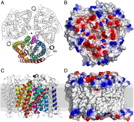
In humans, NH(3) transport across cell membranes is facilitated by the Rh (rhesus) family of proteins. Human Rh C glycoprotein (RhCG) forms a trimeric complex that plays an essential role in ammonia excretion and renal pH regulation. The X-ray crystallographic structure of human RhCG, determined at 2.1 A resolution, reveals the mechanism of ammonia transport. Each monomer contains 12 transmembrane helices, one more than in the bacterial homologs. Reconstituted into proteoliposomes, RhCG conducts NH(3) to raise internal pH. Models of the erythrocyte Rh complex based on our RhCG structure suggest that the erythrocytic Rh complex is composed of stochastically assembled heterotrimers of RhAG, RhD, and RhCE.


Conflict of interest statement

The authors declare no conflict of interest.

Figures

Fig. 1.
Fig. 1.
Molecular structure of RhCG. (A) Cytoplasmic view of the RhCG with monomer in cartoon representation and complete symmetry-generated biological molecule as ribbon. Threefold symmetry axis indicated by ▴, and • indicates the channel position of each monomer. Transmembrane helices are progressively colored blue to red, indicating succession from N to C terminus. (B) Cytoplasmic molecular surface of RhCG colored by electrostatic potential (19) reveals channel apertures surrounded by negative charge (red) with positive charge (blue) at the periphery. (C and D) Representation of A and B, respectively, rotated 90° in membrane (gray) cross-section with cytoplasmic surface up.
Fig. 2.
Fig. 2.
The “M0 lock” in RhCG. (A) Cartoon representation of RhCG rendered in PyMol (20). The side chains of Q19 on M0 and Q101 on M2 are shown as stick representation colored by atom type. The hydrogen bonding between the side chains are indicted by dashed lines. (B) Closer view of the M0 lock with all side chains displayed as sticks and colored by atom type (C: white; O: red; N: blue; S: yellow).
Fig. 3.
Fig. 3.
RhCG NH3 channel and shunt. Partially transparent surface generated by HOLLOW (22) of the channel, voids, and shunt. Select side chains that compose this surface are displayed as sticks. Water molecules within the channel surface displayed as red spheres. All atoms colored by atom type with carbons in white except for the shunt with carbons in green.
Fig. 4.
Fig. 4.
Channel NH3 transport in proteoliposomes. (A) Relative vesicular pH change indicated by CF fluorescence for RhCG at protein to lipid ratio 1∶90 by weight (open blue triangles) and 1∶45 (closed blue triangles) demonstrated protein dependent NH3 permeability relative to empty liposomes (black squares) and a control protein YaaH (red diamonds). (B) Proteoliposomes from the same sample preparations after addition of 275 mM sucrose at t = 0 were monitored for transport of water resulting in decreased vesicular volume, increased CF concentration, and subsequent self-quenching. Data displayed for the exponential fluorescence decay after lag time to reach self-quenching concentration and exponential fit (black lines).
Fig. 5.
Fig. 5.
Phenylalanine gate and polar cluster P3. The partially transparent channel surface and nearby void surfaces are displayed to reveal the waters with observed electron density in the molecular structure. The side chains of Phe gate and P3 residues are shown as sticks colored by atom type. The P3 cluster for each monomer (structure model in white) involves Y254 from the adjacent monomer (labeled with * and in blue).
Fig. 6.
Fig. 6.
Proposed mechanism of acid/ammonium balance in the epithelium of the acid-secreting renal intercalated cells. Channels and pumps are indicated as tubes through the membrane. Substrates are indicated as atomic models. The net flow of respective substrates and diffusion through the membrane are indicated by arrows. Figure is based on the proposed epithelial model for acid-secreting intercalated cells (7, 47, 48).

Similar articles

Cited by

References

    1. Levine P, Stetson RE. An unusual case of intra-group agglutination. J Am Med Assoc. 1939;113:126–127. - PubMed
    1. Colin Y, et al. Genetic basis of the RhD-positive and RhD-negative blood group polymorphism as determined by Southern analysis. Blood. 1991;78:2747–2752. - PubMed
    1. Hartel-Schenk S, Agre P. Mammalian red cell membrane Rh polypeptides are selectively palmitoylated subunits of a macromolecular complex. J Biol Chem. 1992;267:5569–5574. - PubMed
    1. Eyers SA, Ridgwell K, Mawby WJ, Tanner MJ. Topology and organization of human Rh (rhesus) blood group-related polypeptides. J Biol Chem. 1994;269:6417–6423. - PubMed
    1. Marini AM, Urrestarazu A, Beauwens R, Andre B. The Rh (rhesus) blood group polypeptides are related to NH4+ transporters. Trends Biochem Sci. 1997;22:460–461. - PubMed

Publication types

MeSH terms