. 2010 May 25;121(20):2211-20.
doi: 10.1161/CIRCULATIONAHA.109.928796. Epub 2010 May 10.

Mobilized human hematopoietic stem/progenitor cells promote kidney repair after ischemia/reperfusion injury

Affiliations

Mobilized human hematopoietic stem/progenitor cells promote kidney repair after ischemia/reperfusion injury

Bing Li et al. Circulation. .

Abstract

Background: Understanding the mechanisms of repair and regeneration of the kidney after injury is of great interest because there are currently no therapies that promote repair, and kidneys frequently do not repair adequately. We studied the capacity of human CD34(+) hematopoietic stem/progenitor cells (HSPCs) to promote kidney repair and regeneration using an established ischemia/reperfusion injury model in mice, with particular focus on the microvasculature.

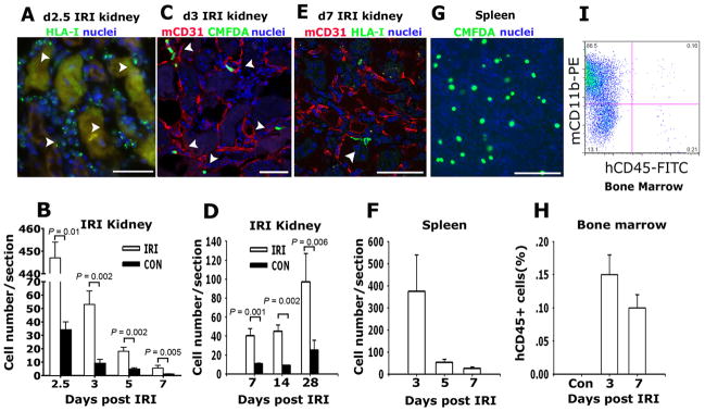
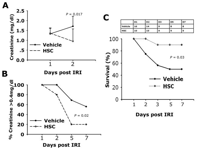
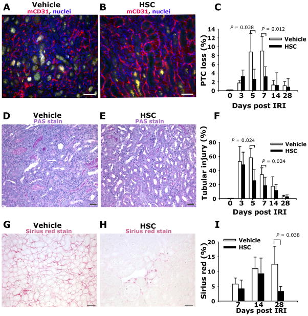
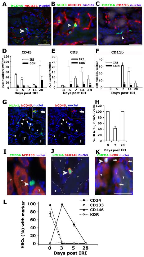
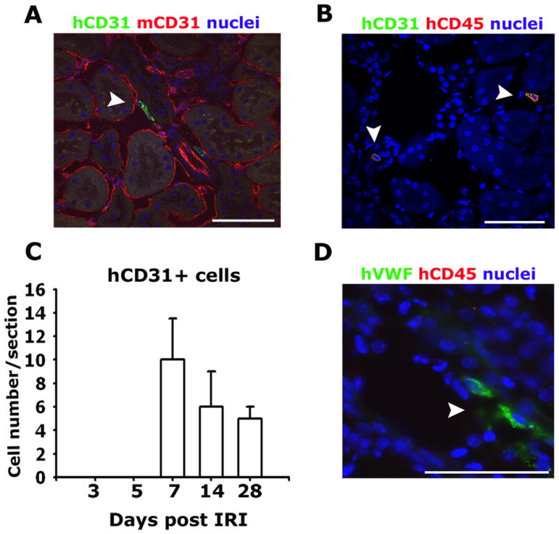
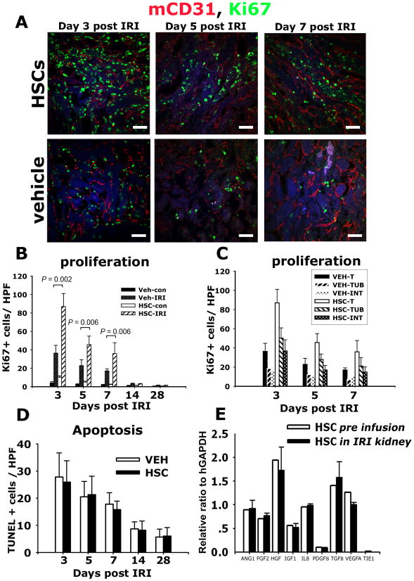
Methods and results: Human HSPCs administered systemically 24 hours after kidney injury were selectively recruited to injured kidneys of immunodeficient mice (Jackson Labs, Bar Harbor, Me) and localized prominently in and around vasculature. This recruitment was associated with enhanced repair of the kidney microvasculature, tubule epithelial cells, enhanced functional recovery, and increased survival. HSPCs recruited to kidney expressed markers consistent with circulating endothelial progenitors and synthesized high levels of proangiogenic cytokines, which promoted proliferation of both endothelial and epithelial cells. Although purified HSPCs acquired endothelial progenitor markers once recruited to the kidney, engraftment of human endothelial cells in the mouse capillary walls was an extremely rare event, indicating that human stem cell mediated renal repair is by paracrine mechanisms rather than replacement of vasculature.

Conclusions: These studies advance human HSPCs as a promising therapeutic strategy for promoting renal repair after injury.


Figures

Figure 1
Figure 1
Human hematopoietic stem cells are recruited to post ischemia reperfusion injury kidneys, spleen and bone marrow of NOD/SCID mice. (A) Photomicrograph of NOD/SCID mouse d2.5 post IRI kidney that received adoptively transferred human HSPCs on d1, showing HLA-I positive cells in the interstitium of the post IRI kidney (arrowheads) between necrotic tubules. (B) Graph indicating the number of human CMFDA+ HSPCs identified in post IRI and control kidneys with time after IRI. (C) Representative confocal image of day 3 post IRI kidney following IV transferred of human HSPCs on d1 post IRI showing CMFDA fluorescing human cells (arrowheads) in peritubular CD31+ capillaries. (D) Graph indicating the number of human HLA class I cells in post IRI and control kidneys on days 7, 14 and 28 after IRI. (E) Representative confocal image detecting human HLA class I (green, arrow) of day 7 post IRI kidney of NOD/SCID mice treated with human HSPCs d1 after IRI. (F) Representative confocal image of CMFDA labeled human HSPCs in spleen 3d following IRI, adoptively transferred 1d after kidney IRI. (G) Graph indicating the number of CMFDA positive cells per section in the spleen on days 3, 5 and 7 after IRI. (H) Graph indicating proportion of human CD45+ cells in mouse bone marrow following adoptive transfer. (I) Representative flow cytometric plot for CD11b (detects mouse and human antigens) and human CD45 of whole bone marrow from d7 post kidney IRI mouse that received adoptively transferred human HSPCs d1 after IRI. Mean ± SD. **P < 0.01 vs. control kidney. (Bars, 50μm, n = 6–10/group)
Figure 2
Figure 2
Adoptive transfer of human HSPCs to NOD/SCID mice following kidney ischemia reperfusion injury decreases mortality and improves kidney function. (A) Plasma creatinine levels on days 1 and 2 following bilateral IRI followed IV injection with PBS (Vehicle, n=16) or 2.5×10⁶ (HSC, n=10) 1day following injury. Data are mean ± SD. (B) Curves showing the proportion of mice with plasma creatinine ≤0.4 mg/dL at each time point following IRI. (C) Survival curves and number at each time point, for mice undergoing bilateral IRI followed IV injection with PBS (vehicle) or 2.5×10⁶ human HSPCs (HSC) 1 day following injury.
Figure 3
Figure 3
Adoptive transfer of Human HSPCs attenuates peritubular capillary loss and reduces tubular epithelial injury following kidney ischemia reperfusion injury. (A–B) Representative images of mouse CD31-labeled peritubular capillaries (PTC) of outer medulla of d7 post IRI kidney that received vehicle (A) or HSPCs (B) on d1 and d2. Note marked PTC loss in (A). (C) Graph showing PTC index for mice following vehicle or HSPCs (n=3 per timepoint). (D–E) Representative light images of PAS stained kidney sections of outer medulla d5 post IRI kidney from mice that received vehicle (D) or HSPCs (E) on d1 and d2. Note prominent debris in severely injured tubules in (D), present to a much lower extent in (E). (F) Graph showing tubular injury index for mice following vehicle or HSPCs (n = 6–10 per timepoint). (G–H) Representative images of Sirius red-stained kidneys d28 post IRI that received either vehicle (G) or HSPCs (H) on d1 and d2 post IRI. (I) Graph showing fibrosis area for mice following vehicle or HSPCs (n = 6–10 per timepoint). Mean ± SD. *P < 0.05, vs. vehicle group. (Bars = 50μm).
Figure 4
Figure 4
Differentiation of human HSPCs in kidneys. Representative confocal image (A) and epifuorescence images (B–C) of kidney outer medulla showing expression of human CD45 (A), human CD3 (B), and absence of CD11b (C) in cells (arrowheads) in d5 post IRI kidneys that received IV injection of HSPCs 1 day following injury. Images are co-labeled (AB) to show mCD31 (red) of the mouse vasculature. Graphs showing the number of human CD45+ (D), human CD3+ (E), and CD11b+; CMFDA+ (F) cells identified in post IRI kidneys and control kidneys. (G–H) Confocal split panel image (G) and graph (H) showing the presence of HLA-I+, hCD45− cells (arrowheads) in the interstitium of the d7 post IRI kidney [T = tubule]. Inset shows a HLA-I+, hCD45+ leukocyte (arrow). (I–K) Representative epifluorescence images of the outer medulla of day 3 post IRI kidney of NOD/SCID mice that received human HSPCs on d1–d2 labeled with CMFDA (arrowheads) co-labeled with antibodies against human CD133 (I), CD146 (J) and KDR (K). Note anti-KDR antibodies also detected mouse endothelium (arrows) [T = tubule, outlined with hatched white line in J]. (L) Graph showing the expression of stem cell/CEP markers by HSPCs prior to (d0) and following recruitment to the post IRI kidney. (Bars = 50μm). Data are mean ± SD. n = 6/timepoint.
Figure 5
Figure 5
Rare human endothelial cells are detected in the kidney after ischemic injury and HSC infusion. (A–B) Confocal images of d28 post IRI kidneys showing the presence of human CD31 expressing cells some of which appear to be integrated into capillaries (arrowhead) (A), but the majority are morphologically monocytic and co-express hCD45 (arrowheads) (B). (C) Graph showing the number of human CD31 expressing cells in the post IRI kidneys with time following adoptive transfer of HSPCs 1day following injury. (D) Specific expression of human von Willebrand factor (vWF) (arrowheads) and not mouse vWF in cells that lack expression of human CD45 in the post IRI kidney (Bar = 50μm).
Figure 6
Figure 6
Human HSPCs generate angiogenic paracrine factors in the kidney after ischemia resperfusion injury that promote parenchymal cell proliferation. (A–C) Confocal images (A) and quantification (B–C) of Ki67+ cells in kidney (B), or kidney tubule (TUB) or interstitial (INT) compartments (C). (D) T.U.N.E.L. positive apoptotic cells in kidney sections post IRI. (E) Relative gene expression compared with GAPDH of pro-angiogenic transcripts in mobilized HSPCs prior to transfer to mice (white) and those purified from post-IRI kidney 48h following transfer to mice. Note that HSPCs recruited to the kidney retain high-level expression of pro-angiogenic transcripts. Mean ± SD.**P < 0.01 n = 6/group. (Bars = 50μm).
Figure 7
Figure 7
Model of functions of HSPCs in repair of the kidney following injury. HSPCs are recruited to the injured kidney where they acquire the CEP marker CD146 and localize within injured capillaries and in the interstitium. Local production of cytokines including Angiopoietins, Vascular endothelial growth factors, hepatocyte growth factor and insulin like growth factors are generated promoting cellular repair by paracrine mechanisms.

References

    1. Ali T, Khan I, Simpson W, Prescott G, Townend J, Smith W, Macleod A. Incidence and outcomes in acute kidney injury: a comprehensive population-based study. J Am Soc Nephrol. 2007;18:1292–1298. - PubMed
    1. Meguid El Nahas A, Bello AK. Chronic kidney disease: the global challenge. Lancet. 2005;365:331–340. - PubMed
    1. Thadhani R, Pascual M, Bonventre JV. Acute renal failure. N Engl J Med. 1996;334:1448–1460. - PubMed
    1. Weiner DE, Tighiouart H, Amin MG, Stark PC, MacLeod B, Griffith JL, Salem DN, Levey AS, Sarnak MJ. Chronic kidney disease as a risk factor for cardiovascular disease and all-cause mortality: a pooled analysis of community-based studies. J Am Soc Nephrol. 2004;15:1307–1315. - PubMed
    1. Sagrinati C, Ronconi E, Lazzeri E, Lasagni L, Romagnani P. Stem-cell approaches for kidney repair: choosing the right cells. Trends Mol Med. 2008;14:277–285. - PubMed

Publication types

MeSH terms