. 2010 Oct;58(10):917-27.
doi: 10.1369/jhc.2010.956243. Epub 2010 May 10.

Quantitative assessment of specificity in immunoelectron microscopy

Affiliations

Quantitative assessment of specificity in immunoelectron microscopy

John Milton Lucocq et al. J Histochem Cytochem. 2010 Oct.

Abstract

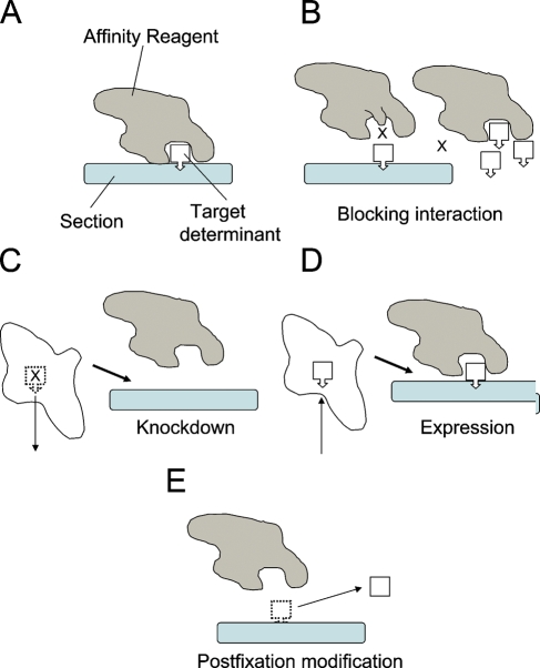
In immunoelectron microscopy (immuno-EM) on ultrathin sections, gold particles are used for localization of molecular components of cells. These particles are countable, and quantitative methods have been established to estimate and evaluate the density and distribution of "raw" gold particle counts from a single uncontrolled labeling experiment. However, these raw counts are composed of two distinct elements: particles that are specific (specific labeling) and particles that are not (nonspecific labeling) for the target component. So far, approaches for assessment of specific labeling and for correction of raw gold particle counts to reveal specific labeling densities and distributions have not attracted much attention. Here, we discuss experimental strategies for determining specificity in immuno-EM, and we present methods for quantitative assessment of (1) the probability that an observed gold particle is specific for the target, (2) the density of specific labeling, and (3) the distribution of specific labeling over a series of compartments. These methods should be of general utility for researchers investigating the distribution of cellular components using on-section immunogold labeling.


Figures

Figure 1
Figure 1
Reagent-based and specimen-based controls. (A) Terminology used in this report. The primary affinity reagent (Affinity Reagent) becomes associated with a target determinant (carried on a target component molecule), which is embedded within the ultrathin section (Section). (B) Reagent-based controls. Left: Interactions of reagent with target determinant can be blocked by modification (in this case mutation) of the binding site, thereby reducing labeling. The affinity reagent is otherwise similar in amino acid sequence, ensuring nonspecific interactions can be preserved. Right: Excess soluble target determinants are introduced into the system and compete for binding of target determinants in the section. In this type of control, effects of the point mutation or competition for binding can be independently verified using biochemical tests for specificity. (C–E) Specimen-based controls by modification of the target component determinant. In C, target determinants are reduced by restricted or deleted expression, which is ideally verified by independent biochemical means. In D, target components carrying target determinants are introduced to the system in this case by inducing expression, although a variety of other methods could be used (see text). Often convenient determinants (such as epitope tags) may be introduced to the target components to facilitate labeling. Preferably, in this type of system, the expression of the endogenous target may need to be suppressed, and the introduced target should be at similar levels to that of endogenous. In E, the determinant is modified chemically before labeling in order to modify the interaction of the affinity reagent with the determinant. In this case, the section has been exposed to an enzyme, which modifies the determinant by cleaving specifically the bond between the determinant and the target component. A good example in this case is the localization of phosphotyrosine (pTyr) using specific antibody. The section is exposed to a phosphatase with specificity for this epitope, and the section is then labeled using the phosphotyrosine antibody. Reduction in the labeling indicates specific labeling for the pTyr.
Figure 2
Figure 2
Assessing specific labeling—simple illustrative example. Microscopic fields were sampled appropriately from ultrathin sections labeled for a membrane antigen—one field from normal expression (NORMAL) condition and one from the reduced expression (REDUCED EXPRESSION) condition. Gold particles are indicated by black dots, and the cell compartment profiles were identified as indicated (for simplicity, nuclear envelope was drawn as a single membrane). Arrays of test lines were overlaid on the micrographs and intersections of one edge of each line with the compartmental membrane profiles were counted. The results are presented in Table 4 and discussed in the Results and Discussion section.

Similar articles

Cited by

References

    1. Abramoff MD, Magelhaes PJ, Ram SJ (2004) Image processing with ImageJ. Biophotonics International 11:36–42
    1. Al-Amoudi A, Chang JJ, Leforestier A, McDowall A, Salamin LM, Norlén LP, Richter K, et al. (2004) Cryo-electron microscopy of vitreous sections. EMBO J 23:3583–3588 - PMC - PubMed
    1. Andjelkovic M, Alessi DR, Meier R, Fernandez A, Lamb NJ, Frech M, Cron P, et al. (1997) Role of translocation in the activation and function of protein kinase B. J Biol Chem 272:31515–31524 - PubMed
    1. Bendayan M (1981) Ultrastructural localization of nuclei acids by the use of enzyme-gold complexes. J Histochem Cytochem 29:531–541 - PubMed
    1. Bendayan M (2000) A review of the potential and versatility of colloidal gold cytochemical labeling for molecular morphology. Biotech Histochem 75:203–242 - PubMed

MeSH terms