. 2010 Jun;120(6):2058-69.
doi: 10.1172/JCI42032. Epub 2010 May 10.

Lnk constrains myeloproliferative diseases in mice

Affiliations

Lnk constrains myeloproliferative diseases in mice

Alexey Bersenev et al. J Clin Invest. 2010 Jun.

Abstract

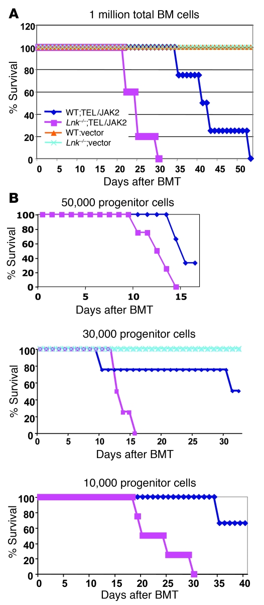
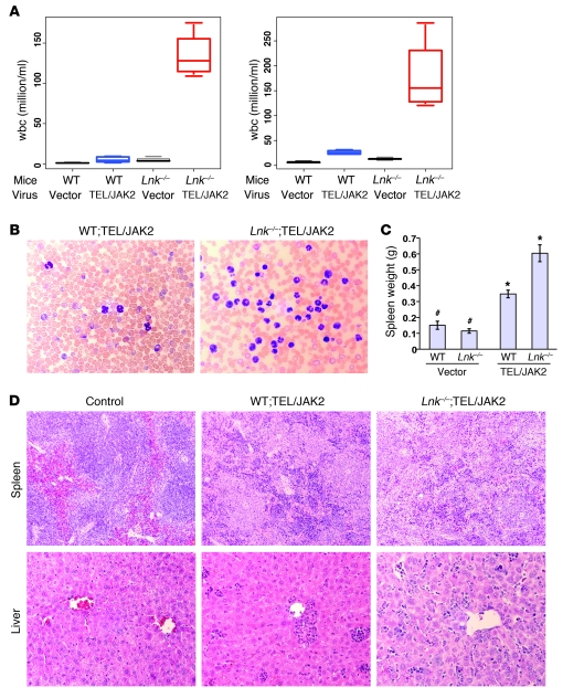
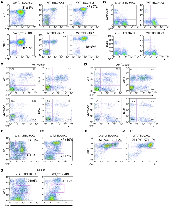
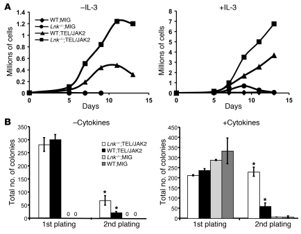
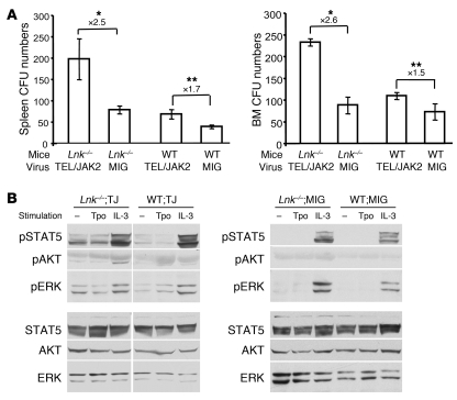
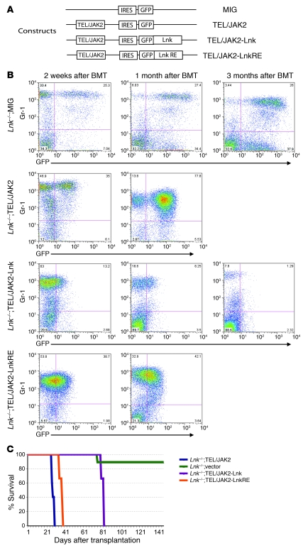
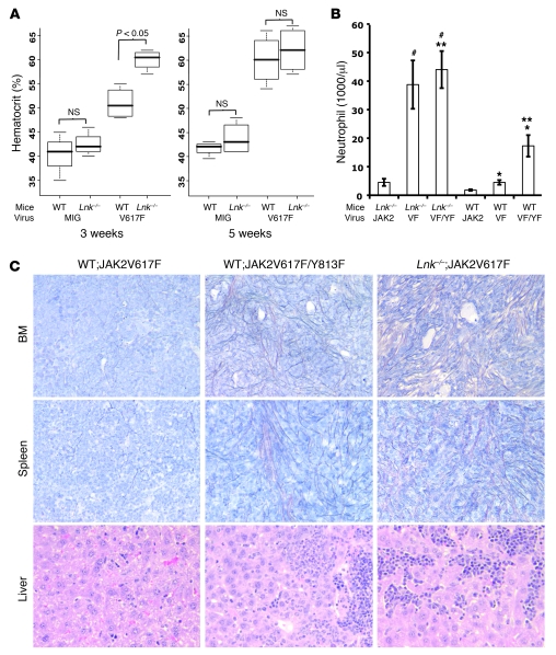
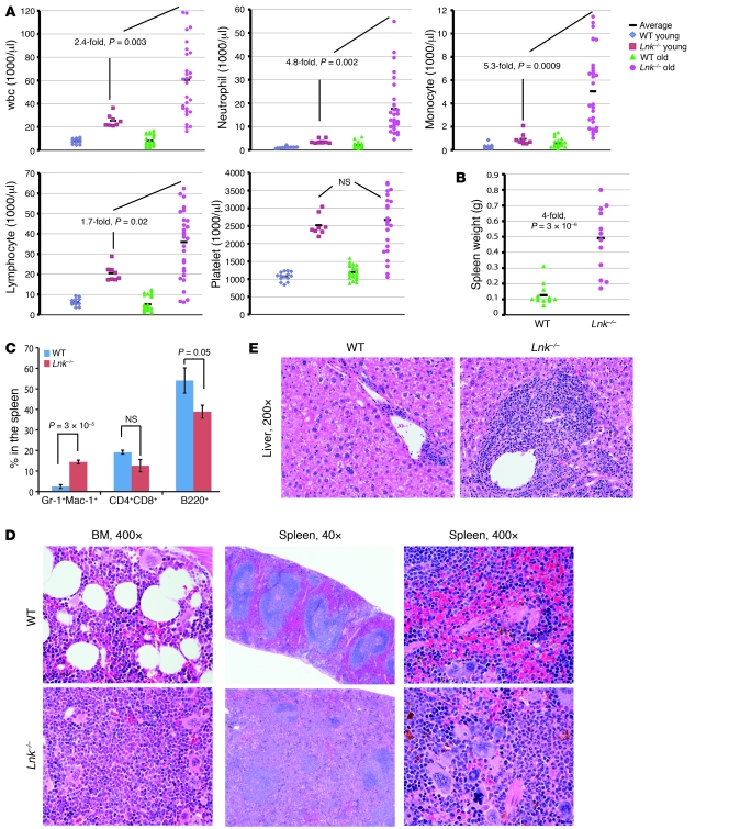
Hematopoietic stem and progenitor cell (HSPC) expansion is regulated by intrinsic signaling pathways activated by cytokines. The intracellular kinase JAK2 plays an essential role in cytokine signaling, and activating mutations in JAK2 are found in a number of hematologic malignancies. We previously demonstrated that lymphocyte adaptor protein (Lnk, also known as Sh2b3) binds JAK2 and attenuates its activity, thereby limiting HSPC expansion. Here we show that loss of Lnk accelerates and exacerbates oncogenic JAK2-induced myeloproliferative diseases (MPDs) in mice. Specifically, Lnk deficiency enhanced cytokine-independent JAK/STAT signaling and augmented the ability of oncogenic JAK2 to expand myeloid progenitors in vitro and in vivo. An activated form of JAK2, unable to bind Lnk, caused greater myeloid expansion than activated JAK2 alone and accelerated myelofibrosis, indicating that Lnk directly inhibits oncogenic JAK2 in constraining MPD development. In addition, Lnk deficiency cooperated with the BCR/ABL oncogene, the product of which does not directly interact with or depend on JAK2 or Lnk, in chronic myeloid leukemia (CML) development, suggesting that Lnk also acts through endogenous pathways to constrain HSPCs. Consistent with this idea, aged Lnk-/- mice spontaneously developed a CML-like MPD. Taken together, our data establish Lnk as a bona fide suppressor of MPD in mice and raise the possibility that Lnk dysfunction contributes to the development of hematologic malignancies in humans.


Figures

Figure 1
Figure 1. Lnk deficiency exacerbates MPD development initiated by TEL/JAK2 in mice.
(A) WT and Lnk–/– BM cells were infected with retroviruses encoding either MIG vector alone or TEL/JAK2, and 1 million total BM cells were transplanted into each irradiated host animal. Kaplan-Meier survival analysis of transplanted mice is shown. P < 0.05, long-rank test comparing WT;TEL/JAK2 and Lnk–/–;TEL/JAK2 groups. n = 5. (B) Lnk deficiency exacerbates CML development initiated by TEL/JAK2 in mice when defined numbers of purified progenitors were transplanted into the hosts. Magnetic bead–enriched Lin progenitor cells from WT and Lnk–/– mice were infected with retroviruses, and equal numbers of WT and Lnk–/– progenitors were transplanted into irradiated host animals. Kaplan-Meier survival analysis of 3 transplants with different progenitor numbers (left, 50,000 progenitor cells; middle, 30,000 progenitor cells; right, 10,000 progenitor cells) is shown. n = 5 in each group of each transplant.
Figure 2
Figure 2. Lnk deficiency leads to an exacerbated neutrophilia and neutrophil infiltration into multiple organs.
(A) The box plot shows wbc measured 2–3 weeks after the transplant (left) and in moribund animals (right). The top and bottom ends of the boxes define the 75th and 25th percentiles, the horizontal lines indicate the medians, and the error bars define the 5th and 95th percentiles. n = 5. P < 0.01 comparing WT;TEL/JAK2 and Lnk–/–;TEL/JAK2 groups. (B) Wright-Giemsa staining of blood smears from WT;TEL/JAK2 and Lnk–/–;TEL/JAK2 mice shows massively increased numbers of myeloid cells at 2–3 weeks. Original magnification, ×400. (C) Spleen weight of transplanted mice (average ± SEM). #P = NS; *P < 0.005. n = 4–6. (D) H&E staining of tissue sections. Original magnification, ×100 (top row, spleen); ×200 (bottom row, liver).
Figure 3
Figure 3. Lnk deficiency leads to an exacerbated neutrophilia, with an expansion of immature myeloid cells in the BM and spleen.
Peripheral blood and tissue lineage chimerism analysis after transplantation was performed using flow cytometry. (A) Myeloid chimerism of Lnk–/–;TEL/JAK2 (left) and WT;TEL/JAK2 (middle) mice 2 weeks after BMT and moribund WT;TEL/JAK2 mice (right). Percentages (average ± SEM) of GFP+ myeloid cells are shown. P < 0.001 comparing WT;TEL/JAK2 and Lnk–/–;TEL/JAK2 groups. (B) Lymphoid chimerism of Lnk–/–;TEL/JAK2 and WT;TEL/JAK2 mice. (C and D) Blood lineage chimerism of WT;vector and Lnk–/–;vector mice 3–4 weeks after BMT. Representative percentages of lineage distributions versus GFP expression are indicated. (EG) Myeloid composition in the BM (E and F) and spleen (G) of WT;TEL/JAK2 and Lnk–/–;TEL/JAK2 mice. (E) The percentage of GFP+Gr-1hi and GFP+Gr-1lo BM cells is quantified (average ± SEM). n = 8. P < 0.01. (F) Mac-1 and Gr-1 expression of GFP+-gated BM cells. The percentage of Mac-1+Gr-1hi mature and Mac-1+Gr-1lo immature myeloid cells is quantified (average ± SEM). n = 8. P < 0.01. (G) The percentage of GFP+Gr-1lo splenic cells (average ± SEM). n = 6. P < 0.05.
Figure 4
Figure 4. Lnk deficiency leads to an enhanced progenitor cell proliferation and self-renewal in vitro.
Lin progenitor cells from WT and Lnk–/– mice were infected with retroviruses encoding either vector alone or TEL/JAK2 and subsequently sorted for GFP positivity. (A) Purified GFP+Lin progenitors were cultured in the absence of cytokine (left) and in the presence of WEHI supernatant containing IL-3 (right). Live cells were enumerated every 2–3 days, and representative growth curves are shown. The graphs show representatives of 5 independent experiments. (B) Purified GFP+Lin progenitors were plated in cytokine-free (M3234) or cytokine-rich (M3434) methylcellulose media (StemCell Technologies Inc.). Eight to ten days later, colonies were enumerated, replated, and scored again another 8–10 days later. The total colony numbers of 1st and 2nd platings are shown (average ± SD). *P < 0.01. n = 4–5.
Figure 5
Figure 5. Lnk deficiency increases the ability of TEL/JAK2 to expand progenitors and enhances cytokine signaling.
Two weeks after BMT, spleen and BM cells from mice reconstituted with WT and Lnk–/– cells expressing MIG or TEL/JAK2 (TJ) were isolated. (A) Spleen and BM cells were plated on M3434 methylcellulose media, and CFU progenitors were quantified at days 8–10. Fold differences (average ± SEM) between indicated groups are shown above the bars. *P < 0.01; **P < 0.05. n = 4. (B) BM cells were starved, then either left unstimulated or stimulated with either 10 ng/ml of Tpo or IL-3 for 10 minutes before lysis. Protein lysates were subjected to Western blot analysis. Representative results of 4 independent experiments are shown.
Figure 6
Figure 6. WT Lnk but not SH2- deficient Lnk can rescue CML development from Lnk–/– cells expressing TEL/JAK2.
(A) Schematic diagram of retroviral constructs used for infection and transplantation. Lin progenitor cells from Lnk–/– mice were infected with retroviruses encoding either MIG, TEL/JAK2, TEL/JAK2-Lnk, or TEL/JAK2-LnkR364E (TEL/JAK2-LnkRE) and transplanted into irradiated recipients. (B) Myeloid chimerisms of reconstituted mice at indicated time points after transplantation. (C) Survival curves of reconstituted mice.
Figure 7
Figure 7. Lnk deficiency accelerates the onset of PV initiated by JAK2V617F and MF progression in mice.
(A) The box plot shows hematocrits measured from mice transplanted with WT and Lnk–/– cells infected with retroviruses (either MIG vector or JAK2V617F) at week 3 and week 5. The top and bottom ends of the boxes define the 75th and 25th percentiles, the horizontal lines indicate the medians, and the error bars define the 5th and 95th percentiles. n = 5; 2-tailed Student’s t tests are shown. (B) Lnk restricts myeloid expansion of JAK2V617F-reconstituted mice. WT and Lnk–/– BM cells were infected with JAK2wt, JAK2V617F, or JAK2V617F/Y813F viruses and transplanted into irradiated recipients. Neutrophil counts in the peripheral blood were analyzed 2 months after transplant (average ± SEM). VF, JAK2V617F; VF/YF, JAK2V617F/Y813F. #P = NS; *P < 0.005; **P < 0.05. (C) Reticulin stainings are shown in the BM and spleen sections (top and middle panels), and H&E stainings are shown for the liver sections (lower panel) of transplanted mice. Original magnification, ×200.
Figure 8
Figure 8. Aged Lnk–/– mice develop a CML-like MPD phenotype.
(A) Peripheral blood cell counts of young and old Lnk–/– mice in comparison with age-matched WT mice. Each symbol represents an individual mouse. Average blood counts are indicated by horizontal lines. Young mice are 2 months of age, and old mice are 12–18 months of age. (B) Spleen weight of old WT and Lnk–/– mice. Average spleen weights are indicated by horizontal lines. (C) Percentage of Gr-1+Mac-1+ myeloid cells, CD4+/CD8+ T cells, and B220+ B cells in the spleen of WT and Lnk–/– mice (average ± SEM). n = 5. Two-tailed t tests were performed, and P values are indicated on the plots. (D and E) H&E stainings of the sections from BM located within the femur, spleen, and liver of aged WT and Lnk–/– mice are shown. Original magnification, ×40 (D, center panel); ×200 (E); ×400 (D, left and right panels).

References

    1. Watowich SS, Wu H, Socolovsky M, Klingmuller U, Constantinescu SN, Lodish HF. Cytokine receptor signal transduction and the control of hematopoietic cell development. Annu Rev Cell Dev Biol. 1996;12:91–128. doi: 10.1146/annurev.cellbio.12.1.91. - DOI - PubMed
    1. Drachman JG, Kaushansky K. Structure and function of the cytokine receptor superfamily. Curr Opin Hematol. 1995;2(1):22–28. doi: 10.1097/00062752-199502010-00004. - DOI - PubMed
    1. Schlessinger J, Lemmon MA. SH2 and PTB domains in tyrosine kinase signaling. Sci STKE. 2003;2003(191):RE12. - PubMed
    1. Rudd CE. Lnk adaptor: novel negative regulator of B cell lymphopoiesis. Sci STKE. 2001;2001(85):pe1. - PubMed
    1. Takaki S, et al. Control of B cell production by the adaptor protein lnk. Definition Of a conserved family of signal-modulating proteins. Immunity. 2000;13(5):599–609. - PMC - PubMed

Publication types