Review
. 2010 Jul;104(1):23-9.
doi: 10.1160/TH09-07-0507. Epub 2010 May 10.

Pluripotency of human embryonic and induced pluripotent stem cells for cardiac and vascular regeneration

Affiliations
Review

Pluripotency of human embryonic and induced pluripotent stem cells for cardiac and vascular regeneration

Kenneth R Boheler. Thromb Haemost. 2010 Jul.

Abstract

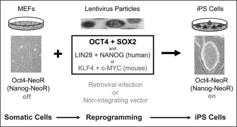
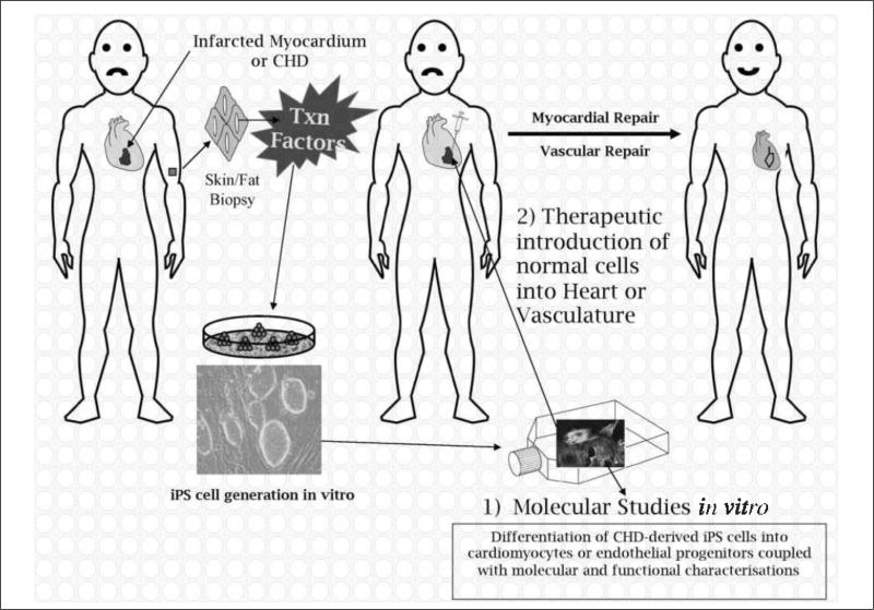
Cardiac and vascular abnormalities and disease syndromes are major causes of death both during human development and with aging. To identify the cause of congenital defects and to combat this epidemic in the aging population, new models must be created for scientific investigation and new therapies must be developed. Recent advances in pluripotent stem cell biology offer renewed hope for tackling these problems. Of particular importance has been the creation of induced pluripotent (iPS) cells from adult tissues and organs through the forced expression of two to four transcription factors. Moreover, iPS cells, which are phenotypically indistinguishable from embryonic stem (ES) cells, can be generated from any patient. This unique capacity when coupled with samples from patients who have congenital and genetic defects of unknown aetiology should permit the creation of new model systems that foment scientific investigation. Moreover, creation of patient-specific cells should overcome many of the immunological limitations that currently impede therapeutic applications associated with other pluripotent stem cells and their derivatives.The aims of this paper will be to discuss cardiac and vascular diseases and show how iPS cells may be employed to overcome some of the most significant scientific and clinical hurdles facing this field.


Figures

Figure 1
Figure 1. Schematic illustration of how mouse (and human) somatic cells can be reprogrammed to pluripotent iPS cells
Reprogramming takes place through a series of time-dependent steps. The first is the introduction of pluripotency transcription factors into somatic cells, which lead to the down-regulation of lineage-specific genes. Markers like endogenous alkaline phosphatase activity subsequently increase followed by SSEA1 up-regulation. At the completion of the reprogramming process, endogenous oct4 and nanog are expressed, which, when targeted to activate antibiotic (e.g. NeoR) selection, can be employed to isolate fully reprogrammed cells. MEFs, mouse embryonic fibroblasts; NeoR, neomycin gene cassette that confers resistance to G418.
Figure 2
Figure 2. Developmental markers in the selection of subsets of mesodermally-derived cells
This schema illustrates the use of developmental markers in the identification of subsets of mesodermally derived cells (smooth muscle cells, cardiac muscle cells, haematopoietic cells, endothelial cells) that can be isolated from differentiating pluripotent cells. Selected developmental markers are indicated and additional lineage specificity is as follows: haematopoietic (Flt1, Scl/Tal1 positive), endothelial (Flk-1-, Flt-1-, Tie-2-positive) and cardiac lineages (Nkx2.5 and Isl1-positive).
Figure 3
Figure 3. Schematic illustration of the generation of iPS cells and how these cells can theoretically be employed to study 1) molecular mechanisms in vitro or 2) used for therapeutic interventions in humans
In the case of myocardial infarctions (or vascular defects), iPS cells could be generated from a patient, differentiated to cardiomyocytes (or endothelial cells) and reintroduced into the damaged tissue to elicit repair. Where iPS cells are produced from patients with CHDs of unknown aetiology, the aim would be to determine the cause of the disease in vitro, and not to produce iPS cells for repair, unless the genetic defect could be repaired in vitro prior to introduction of corrected cells back into the host (see Therapeutic applications for details). CHD, congenital heart defect; Txn factors, transcription factors (Oct4, Sox2, Nanog, Lin28).

References

    1. Pierpont ME, Basson CT, Benson DW, et al. Genetic basis for congenital heart defects: Current knowledge – A scientific statement from the American heart association congenital cardiac defects committee, council on cardiovascular disease in the young. Circulation. 2007;115:3015–3038. - PubMed
    1. Najjar SS, Scuteri A, Lakatta EG. Arterial aging – Is it an immutable cardiovascular risk factor? Hypertension. 2005;46:454–462. - PubMed
    1. Lakatta EG, Wang MY, Najjar SS. Arterial aging and subclinical arterial disease are fundamentally intertwined at macroscopic and molecular levels. Med Clin North Am. 2009;93:583–604. - PMC - PubMed
    1. Chantler PD, Lakatta EG, Najjar SS. Arterial-ventricular coupling: mechanistic insights into cardiovascular performance at rest and during exercise. J Appl Physiol. 2008;105:1342–1351. - PMC - PubMed
    1. Steffens S, Montecucco F, Mach F. The inflammatory response as a target to reduce myocardial ischaemia and reperfusion injury. Thromb Haemost. 2009;102:240–247. - PubMed

Publication types

MeSH terms